Nisha Aji Kankana, Hafizi Sina, Da Silva Tania, Kiang Michael, Rusjan Pablo M, Weickert Cynthia Shannon, Mizrahi Romina
Department of Pharmacology & Toxicology, University of Toronto, Toronto, Ontario, Canada.
Research Imaging Centre, Centre for Addiction and Mental Health, Toronto, Ontario, Canada.
Brain Behav Immun Health. 2023 May 13;30:100636. doi: 10.1016/j.bbih.2023.100636. eCollection 2023 Jul.
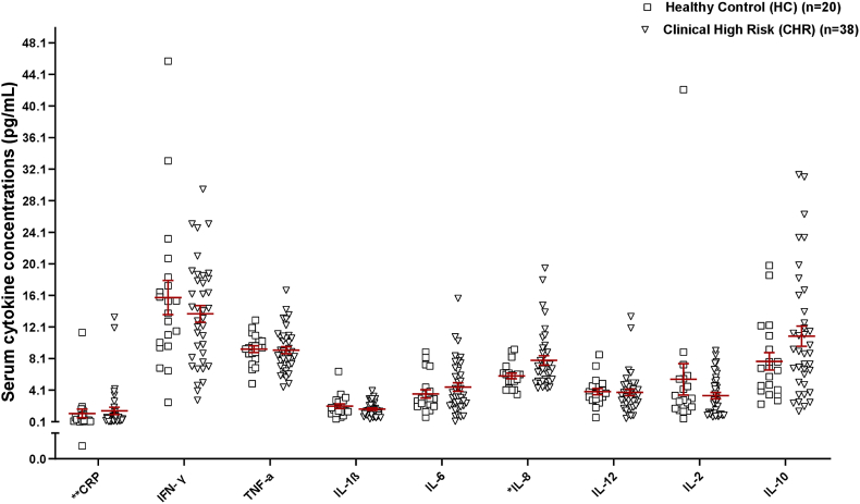
Neuroinflammatory events prior to the diagnosis of schizophrenia may play a role in transition to illness. To date only one study has investigated this association between peripheral proinflammatory cytokines and brain markers of inflammation (e.g., mitochondrial 18 kDa translocator protein, TSPO) in schizophrenia, but none in its putative prodrome. In this study, we primarily aimed to (Barron et al., 2017) test study group (clinical high-risk (CHR) and healthy controls) differences in peripheral inflammatory markers and test for any associations with symptom measures, (Hafizi et al., 2017a) investigate the interaction between brain TSPO levels (dorsolateral prefrontal cortex (DLPFC) and hippocampus) and peripheral inflammatory clusters (entire cohort and (CHR) group independently) within a relatively large group of individuals at CHR for psychosis (N = 38) and healthy controls (N = 20). Participants underwent structural brain magnetic resonance imaging (MRI) and TSPO [F]FEPPA positron emission tomography (PET) scans. Serum samples were assessed for peripheral inflammatory markers (i.e., CRP and interleukins). For exploratory analysis, we aimed to examine cluster differences for symptom measures and identify independent peripheral predictors of brain TSPO expression. Here, we report increased IL-8 levels that are positively correlated with prodromal general symptom severity and showed trend-level association with apathy in CHR. We identified distinct inflammatory clusters characterized by inflammatory markers (IL-1 β, IL-2, IFN-γ) that were comparable between entire cohort and CHR. TSPO levels did not differ between inflammatory clusters (entire cohort or CHR). Finally, we show that CRP, IL-1 β, TNF-α, and IFN-γ levels were the independent peripheral predictors of brain TSPO expression. Thus, alterations in brain TSPO expression in response to inflammatory processes are not evident in CHR. Taken together, clustering by inflammatory status is a promising strategy to characterize the interaction between brain TSPO and peripheral markers of inflammation.
精神分裂症诊断之前的神经炎症事件可能在疾病转变过程中起作用。迄今为止,仅有一项研究调查了精神分裂症患者外周促炎细胞因子与大脑炎症标志物(如线粒体18 kDa转位蛋白,TSPO)之间的这种关联,但尚无研究涉及其假定的前驱期。在本研究中,我们主要旨在(巴伦等人,2017年)测试研究组(临床高危(CHR)和健康对照)在外周炎症标志物方面的差异,并测试与症状指标的任何关联,(哈菲齐等人,2017a)在一组相对较大的精神病临床高危个体(N = 38)和健康对照(N = 20)中,独立研究大脑TSPO水平(背外侧前额叶皮层(DLPFC)和海马体)与外周炎症簇(整个队列和(CHR)组)之间的相互作用。参与者接受了脑部结构磁共振成像(MRI)和TSPO [F]FEPPA正电子发射断层扫描(PET)。评估血清样本中的外周炎症标志物(即CRP和白细胞介素)。为了进行探索性分析,我们旨在检查症状指标的簇差异,并确定大脑TSPO表达的独立外周预测因子。在此,我们报告IL-8水平升高,其与前驱期一般症状严重程度呈正相关,并在CHR中与冷漠呈趋势水平关联。我们确定了以炎症标志物(IL-1β、IL-2、IFN-γ)为特征的不同炎症簇,在整个队列和CHR之间具有可比性。炎症簇(整个队列或CHR)之间的TSPO水平没有差异。最后,我们表明CRP、IL-1β、TNF-α和IFN-γ水平是大脑TSPO表达的独立外周预测因子。因此,在CHR中,大脑TSPO表达因炎症过程而发生的改变并不明显。综上所述,按炎症状态聚类是一种很有前景的策略,可用于表征大脑TSPO与外周炎症标志物之间的相互作用。