Laboratory of Immunoendocrinology, Department of Experimental Neuroendocrinology, Maj Institute of Pharmacology, Polish Academy of Sciences, 12 Smętna St., 31-343 Kraków, Poland.
Department of Biophysical Microstructures, Institute of Nuclear Physics, Polish Academy of Sciences, 152 Radzikowskiego St., 31-342 Kraków, Poland.
Cells. 2023 May 24;12(11):1465. doi: 10.3390/cells12111465.
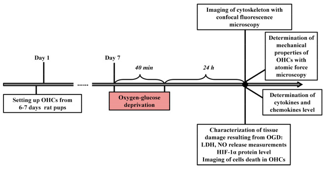
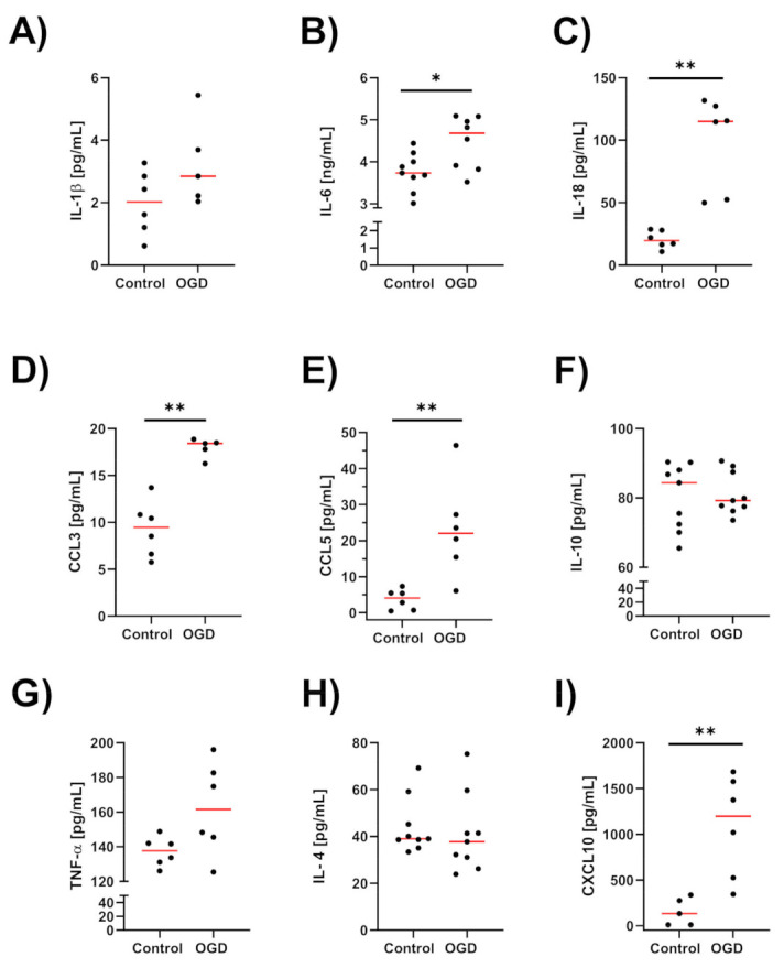
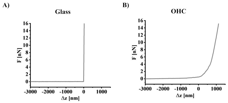
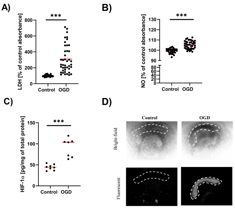
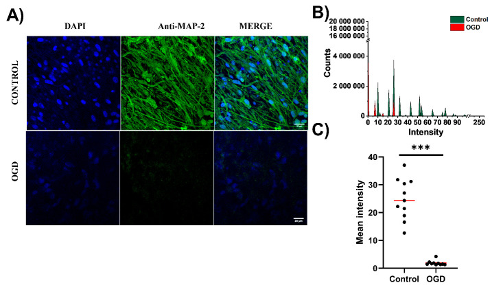
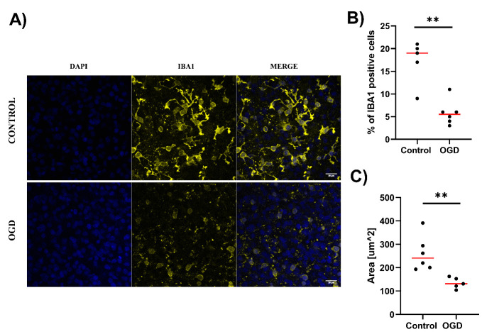
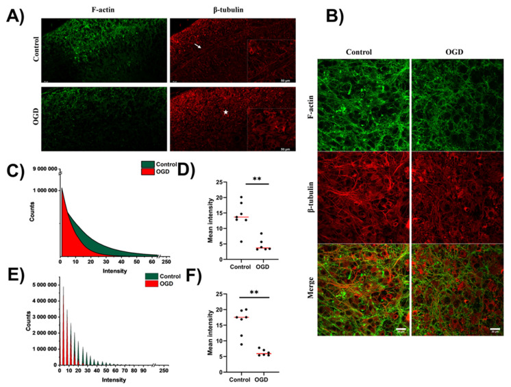
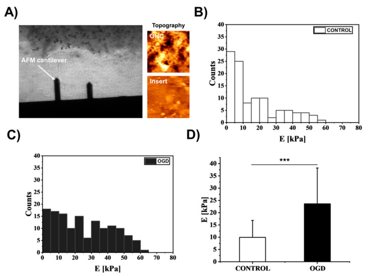
Ischaemic stroke is characterized by a sudden loss of blood circulation to an area of the brain, resulting in a corresponding loss of neurologic function. As a result of this process, neurons in the ischaemic core are deprived of oxygen and trophic substances and are consequently destroyed. Tissue damage in brain ischaemia results from a complex pathophysiological cascade comprising various distinct pathological events. Ischaemia leads to brain damage by stimulating many processes, such as excitotoxicity, oxidative stress, inflammation, acidotoxicity, and apoptosis. Nevertheless, less attention has been given to biophysical factors, including the organization of the cytoskeleton and the mechanical properties of cells. Therefore, in the present study, we sought to evaluate whether the oxygen-glucose deprivation (OGD) procedure, which is a commonly accepted experimental model of ischaemia, could affect cytoskeleton organization and the paracrine immune response. The abovementioned aspects were examined ex vivo in organotypic hippocampal cultures (OHCs) subjected to the OGD procedure. We measured cell death/viability, nitric oxide (NO) release, and hypoxia-inducible factor 1α (HIF-1α) levels. Next, the impact of the OGD procedure on cytoskeletal organization was evaluated using combined confocal fluorescence microscopy (CFM) and atomic force microscopy (AFM). Concurrently, to find whether there is a correlation between biophysical properties and the immune response, we examined the impact of OGD on the levels of crucial ischaemia cytokines (IL-1β, IL-6, IL-18, TNF-α, IL-10, IL-4) and chemokines (CCL3, CCL5, CXCL10) in OHCs and calculated Pearsons' and Spearman's rank correlation coefficients. The results of the current study demonstrated that the OGD procedure intensified cell death and nitric oxide release and led to the potentiation of HIF-1α release in OHCs. Moreover, we presented significant disturbances in the organization of the cytoskeleton (actin fibers, microtubular network) and cytoskeleton-associated protein 2 (MAP-2), which is a neuronal marker. Simultaneously, our study provided new evidence that the OGD procedure leads to the stiffening of OHCs and a malfunction in immune homeostasis. A negative linear correlation between tissue stiffness and branched IBA1 positive cells after the OGD procedure suggests the pro-inflammatory polarization of microglia. Moreover, the negative correlation of pro- and positive anti-inflammatory factors with actin fibers density indicates an opposing effect of the immune mediators on the rearrangement of cytoskeleton induced by OGD procedure in OHCs. Our study constitutes a basis for further research and provides a rationale for integrating biomechanical and biochemical methods in studying the pathomechanism of stroke-related brain damage. Furthermore, presented data pointed out the interesting direction of proof-of-concept studies, in which follow-up may establish new targets for brain ischemia therapy.
缺血性中风的特征是大脑区域突然失去血液循环,导致相应的神经功能丧失。由于这个过程,缺血核心中的神经元会被剥夺氧气和营养物质,因此会被破坏。脑缺血组织损伤是由多种不同病理事件组成的复杂病理生理级联反应的结果。缺血通过刺激兴奋性毒性、氧化应激、炎症、酸中毒和细胞凋亡等多种过程导致脑损伤。然而,人们对包括细胞骨架组织和细胞力学特性在内的生物物理因素关注较少。因此,在本研究中,我们试图评估氧葡萄糖剥夺(OGD)程序是否会影响细胞骨架组织的排列和旁分泌免疫反应,OGD 程序是一种公认的缺血模型。在接受 OGD 程序的器官型海马培养物(OHC)中,我们从体外评估了上述方面。我们测量了细胞死亡/活力、一氧化氮(NO)释放和缺氧诱导因子 1α(HIF-1α)水平。接下来,我们使用共聚焦荧光显微镜(CFM)和原子力显微镜(AFM)评估了 OGD 程序对细胞骨架组织排列的影响。同时,为了确定生物物理特性与免疫反应之间是否存在相关性,我们研究了 OGD 对 OHC 中关键缺血细胞因子(IL-1β、IL-6、IL-18、TNF-α、IL-10、IL-4)和趋化因子(CCL3、CCL5、CXCL10)水平的影响,并计算了 Pearson 和 Spearman 秩相关系数。本研究结果表明,OGD 程序加剧了细胞死亡和一氧化氮释放,并导致 OHC 中 HIF-1α释放增强。此外,我们发现细胞骨架组织(肌动蛋白纤维、微管网络)和细胞骨架相关蛋白 2(MAP-2)的排列出现了明显的紊乱,MAP-2 是一种神经元标志物。同时,我们的研究提供了新的证据,表明 OGD 程序导致 OHC 变硬和免疫稳态功能障碍。OGD 程序后组织硬度与分支 IBA1 阳性细胞之间呈负线性相关,提示小胶质细胞呈炎症前极化。此外,促炎和抗炎因子与肌动蛋白纤维密度呈负相关,表明免疫介质对 OHC 中 OGD 程序诱导的细胞骨架重排具有相反的作用。我们的研究为进一步的研究提供了基础,并为整合生物力学和生化方法研究与中风相关的脑损伤的发病机制提供了依据。此外,所提供的数据指出了一个有趣的概念验证研究方向,后续研究可能会为脑缺血治疗建立新的靶点。