Department of Nephrology, Kameda Medical Center, 929 Higashi-cho, Kamogawa 296-8602, Chiba, Japan.
Department of Nephrology and Blood Purification, Tokyo Medical University Hachioji Medical Center, 1163, Tate-machi, Hachioji 193-0998, Tokyo, Japan.
Int J Mol Sci. 2023 May 27;24(11):9369. doi: 10.3390/ijms24119369.
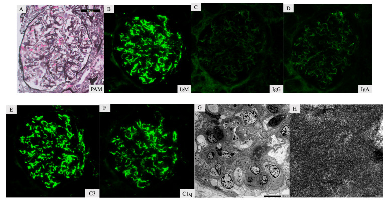
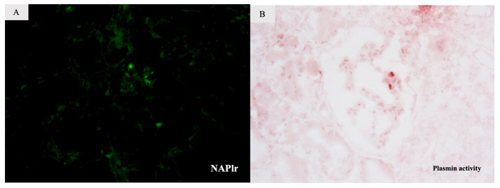
In this rare case of infection-related cryoglobulinemic glomerulonephritis with infective endocarditis, a 78-year-old male presented with an acute onset of fever and rapidly progressive glomerulonephritis. His blood culture results were positive for , and transesophageal echocardiography showed vegetation. He was diagnosed with endocarditis. His serum immunoglobulin M, IgM-cryoglobulin, and proteinase-3-anti-neutrophil cytoplasmic antibody levels were elevated, and his serum complement 3 (C3) and C4 levels were decreased. Renal biopsy results showed endocapillary proliferation, mesangial cell proliferation, and no necrotizing lesions on light microscopy, with strong positive staining for IgM, C3, and C1q in the capillary wall. Electron microscopy showed deposits in the mesangial area in the form of fibrous structures without any humps. Histological examination confirmed a diagnosis of cryoglobulinemic glomerulonephritis. Further examination showed the presence of serum anti-factor B antibodies and positive staining for nephritis-associated plasmin receptor and plasmin activity in the glomeruli, suggesting infective endocarditis-induced cryoglobulinemic glomerulonephritis.
在这例罕见的感染相关性冷球蛋白血症性肾小球肾炎合并感染性心内膜炎中,一名 78 岁男性以发热和快速进行性肾小球肾炎急性起病。他的血培养结果呈阳性,经食管超声心动图检查发现赘生物。他被诊断为心内膜炎。他的血清免疫球蛋白 M、IgM-冷球蛋白和蛋白酶 3-抗中性粒细胞胞质抗体水平升高,血清补体 3(C3)和 C4 水平降低。肾活检结果显示,光镜下可见毛细血管内增生、系膜细胞增生,无坏死病变,毛细血管壁 IgM、C3 和 C1q 强阳性染色。电子显微镜显示在系膜区以纤维状结构形式存在的沉积物,无驼峰。组织学检查证实为冷球蛋白血症性肾小球肾炎。进一步检查显示血清抗因子 B 抗体存在,肾小球中肾炎相关纤溶酶受体和纤溶酶活性阳性染色,提示感染性心内膜炎引起的冷球蛋白血症性肾小球肾炎。