Department of Ophthalmology, Faculty of Medicine and Graduate School of Medicine, Hokkaido University, N-15, W-7, Kita-Ku, Sapporo, 060-8638, Japan.
BMC Ophthalmol. 2023 Jun 13;23(1):270. doi: 10.1186/s12886-023-03026-9.
Metastatic choroidal tumors are hematogenous intraocular metastases of malignant tumors in systemic organs; however, the details of choroidal circulation and morphological changes in the choroid are unknown. The aim of this study is to present a case of metastatic choroidal tumor and examine laser speckle flowgraphy (LSFG)-based choroidal circulation and central choroidal thickness (CCT) before and after chemoradiotherapy.
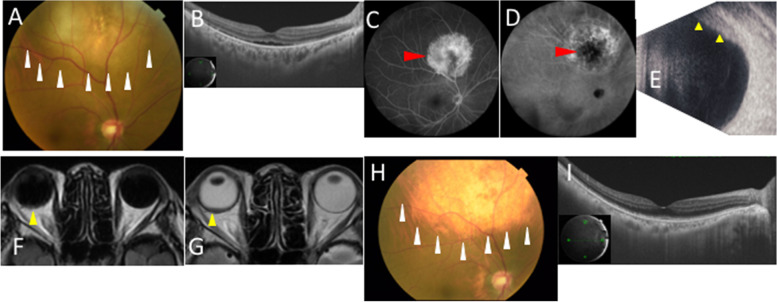
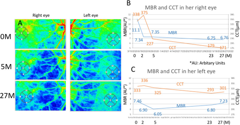
A 66-year-old woman with a medical history of breast cancer 16 years ago was referred to our department struggling with blurred vision in her right eye. At the time of initial examination, her best-corrected visual acuity (BCVA) was 0.4 oculus dexter (OD) and 0.9 oculus sinister. Fundus revealed a yellowish-white choroidal elevated lesion measuring 8 papillary diameters with serous retinal detachment (SRD) in the posterior pole. Fluorescein angiography showed diffuse hyperfluorescence and fluorescent leakage due to SRD, and indocyanine green angiography demonstrated no abnormalities in the macula but hypofluorescence in the center of the tumor. Based on these clinical findings, she was diagnosed with metastatic choroidal tumor. After chemoradiotherapy, the metastatic choroidal tumor became scarred, and SRD disappeared. The rate of changes in macular blood flows assessed by mean blur rate on LSFG and CCT of her right eye were 33.8 and 32.8% decrease at 5 months after the initial visit, respectively. BCVA was 0.5 OD 27 months after the initial examination.
Chemoradiotherapy resulted in regression of the metastatic choroidal tumor and disappearance of SRD, with a decrease in central choroidal blood flow and CCT. The choroidal blood flow on LSFG could reflect an increased oxygen demand by cancer cells invading the choroid and substantial blood supply.
转移性脉络膜肿瘤是全身系统器官恶性肿瘤血行性眼内转移;然而,脉络膜循环的细节和脉络膜形态变化尚不清楚。本研究旨在报道 1 例转移性脉络膜肿瘤病例,并检查化疗和放疗前后基于激光散斑血流图(LSFG)的脉络膜循环和中央脉络膜厚度(CCT)。
一位 66 岁女性,16 年前患有乳腺癌,因右眼视力模糊就诊于我科。初次检查时,她的右眼最佳矫正视力(BCVA)为 0.4 视敏度(OD)和 0.9 视敏度(OS)。眼底检查发现后极部有一个 8 个视乳头直径大小的黄白色脉络膜隆起性病变,伴有浆液性视网膜脱离(SRD)。荧光素血管造影显示弥漫性高荧光和 SRD 荧光渗漏,吲哚菁绿血管造影显示黄斑无异常,但肿瘤中心呈低荧光。根据这些临床发现,她被诊断为转移性脉络膜肿瘤。化疗和放疗后,转移性脉络膜肿瘤发生瘢痕化,SRD 消失。LSFG 评估的黄斑血流变化率和右眼 CCT 的平均模糊率分别为初始就诊后 5 个月时 33.8%和 32.8%的下降。初始检查后 27 个月时,右眼 BCVA 为 0.5 OD。
化疗和放疗导致转移性脉络膜肿瘤消退和 SRD 消失,中央脉络膜血流和 CCT 减少。LSFG 上的脉络膜血流可以反映侵袭脉络膜的癌细胞增加的氧气需求和大量的血液供应。