Langley Christelle, Masuda Naoki, Godwin Simon, De Marco Giovanni, Smith Angela Davies, Jones Rosemary, Bruce Jared, Thai Ngoc Jade
CRIC Bristol, Bristol Medical School, University of Bristol, Bristol, United Kingdom.
Department of Psychiatry, University of Cambridge, Cambridge, United Kingdom.
Front Neurosci. 2023 Jun 2;17:1194859. doi: 10.3389/fnins.2023.1194859. eCollection 2023.
Central fatigue is one of the most common symptoms in multiple sclerosis (MS). It has a profound impact on quality of life and a negative effect on cognition. Despite its widespread impact, fatigue is poorly understood and very difficult to measure. Whilst the basal ganglia has been implicated in fatigue the nature of its role and involvement with fatigue is still unclear. The aim of the present study was to establish the role of the basal ganglia in MS fatigue using functional connectivity measures.
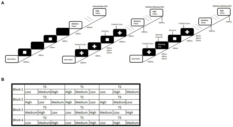
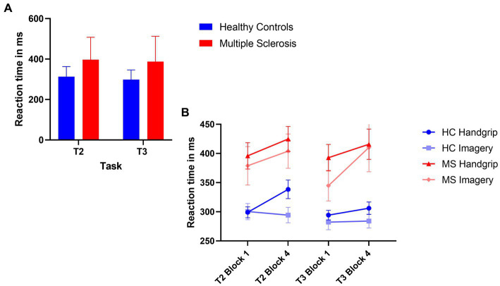
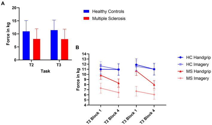
The present study examined the functional connectivity (FC) of the basal ganglia in a functional MRI study with 40 female participants with MS (mean age = 49.98 (SD = 9.65) years) and 40 female age-matched (mean age = 49.95 (SD = 9.59) years) healthy controls (HC). To measure fatigue the study employed the subjective self-report Fatigue Severity Scale and a performance measure of cognitive fatigue using an alertness-motor paradigm. To distinguish physical and central fatigue force measurements were also recorded.
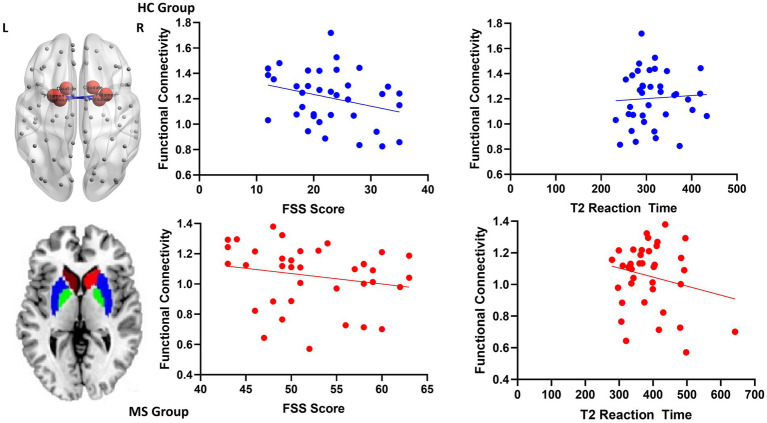
The results suggest that decreased local FC within the basal ganglia plays a key role in cognitive fatigue in MS. Increased global FC between the basal ganglia and the cortex may sub serve a compensatory mechanism to reduce the impact of fatigue in MS.
The current study is the first to show that basal ganglia functional connectivity is associated with both subjective and objective fatigue in MS. In addition, the local FC of the basal ganglia during fatigue inducing tasks could provide a neurophysiological biomarker of fatigue.
中枢性疲劳是多发性硬化症(MS)最常见的症状之一。它对生活质量有深远影响,对认知有负面影响。尽管其影响广泛,但疲劳的机制尚不清楚且很难测量。虽然基底神经节与疲劳有关,但其作用的本质以及与疲劳的关联仍不明确。本研究的目的是使用功能连接测量方法来确定基底神经节在MS疲劳中的作用。
本研究在一项功能磁共振成像研究中,对40名患有MS的女性参与者(平均年龄=49.98(标准差=9.65)岁)和40名年龄匹配的女性健康对照者(HC,平均年龄=49.95(标准差=9.59)岁)的基底神经节功能连接(FC)进行了检查。为了测量疲劳,该研究采用了主观自我报告的疲劳严重程度量表以及使用警觉-运动范式的认知疲劳表现测量方法。为了区分身体疲劳和中枢性疲劳,还记录了力量测量值。
结果表明,基底神经节内局部FC的降低在MS的认知疲劳中起关键作用。基底神经节与皮层之间整体FC的增加可能有助于一种补偿机制,以减少MS中疲劳的影响。
本研究首次表明,基底神经节功能连接与MS中的主观和客观疲劳均相关。此外,疲劳诱导任务期间基底神经节的局部FC可为疲劳提供一种神经生理生物标志物。