Yang Jaehun, Rhu Jinsoo, Kwon Jieun, Choi Gyu-Seong, Kim Jong Man, Jeong Woo Kyoung, Joh Jae-Won
Department of Surgery, Samsung Medical Center, Sungkyunkwan University School of Medicine, Seoul, Korea.
Department of Surgery, Gil Medical Center, Gachon University College of Medicine, Incheon, Korea.
Ann Surg Treat Res. 2023 Jun;104(6):348-357. doi: 10.4174/astr.2023.104.6.348. Epub 2023 Jun 7.
This study evaluated the clinical implication of hepatic venous territory mapping in living donor liver transplantation.
Living donor liver transplantations performed using right graft since 2017 were included. Hepatic venous volume mapping was started in 2019. Risk factors for graft failure and overall survival were analyzed. Analysis for factors related to occlusion of reconstructed vein was performed.
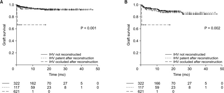
Among 445 patients included, 213 underwent hepatic venous mapping. Hepatic venous mapping itself was not a significant factor for graft (hazard ratio [HR], 0.958; 95% confidence interval [CI], 0.441-2.082; P = 0.913) and overall survival (HR, 0.627; 95% CI, 0.315-1.247; P = 0.183). Inferior hepatic vein occlusion was a significant risk factor for both graft survival (HR, 8.795; 95% CI, 1.628-47.523; P = 0.012) and overall survival (HR, 11.13; 95% CI, 2.460-50.300; P = 0.002). In a subgroup with middle hepatic vein reconstruction, occlusion was a significant risk factor for overall survival (HR, 3.289; 95% CI, 1.304-8.296; P = 0.012). In patients with middle hepatic vein reconstruction whose venous territory volumes were measured, right anterior volume of ≥300 cm was protective for vein occlusion (OR, 0.317; 95% CI, 0.152-0.662; P = 0.002). In patients with V5 reconstruction, V5 volume of ≥150 cm was protective for vein occlusion (OR, 0.253; 95% CI, 0.087-0.734; P = 0.011).
Inferior and middle hepatic vein reconstruction has significant impact on clinical outcome. Hepatic venous territory mapping can provide an objective measure for successful reconstruction of venous branches.
本研究评估了肝静脉区域测绘在活体肝移植中的临床意义。
纳入2017年以来采用右半肝移植物进行的活体肝移植病例。肝静脉容积测绘始于2019年。分析移植物失败和总体生存的危险因素。对与重建静脉闭塞相关的因素进行分析。
在纳入的445例患者中,213例进行了肝静脉测绘。肝静脉测绘本身对移植物(风险比[HR],0.958;95%置信区间[CI],0.441 - 2.082;P = 0.913)和总体生存(HR,0.627;95% CI,0.315 - 1.247;P = 0.183)并非显著因素。肝下静脉闭塞是移植物生存(HR,8.795;95% CI,1.628 - 47.523;P = 0.012)和总体生存(HR,11.13;95% CI,2.460 - 50.300;P = 0.002)的显著危险因素。在肝中静脉重建的亚组中,闭塞是总体生存的显著危险因素(HR,3.289;95% CI,1.304 - 8.296;P = 0.012)。在测量了静脉区域容积的肝中静脉重建患者中,右前叶容积≥300 cm对静脉闭塞具有保护作用(比值比[OR],0.317;95% CI,0.152 - 0.662;P = 0.002)。在V5重建患者中,V5容积≥150 cm对静脉闭塞具有保护作用(OR,0.253;95% CI,0.087 - 0.734;P = 0.011)。
肝下和肝中静脉重建对临床结局有显著影响。肝静脉区域测绘可为静脉分支的成功重建提供客观测量方法。