Department of Ophthalmology, Wuhan Aier eye hospital, Wuhan University, Wuhan, Hubei, China.
BMC Ophthalmol. 2023 Jun 20;23(1):283. doi: 10.1186/s12886-023-03042-9.
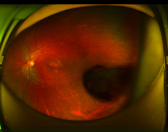
Posterior lenticonus is an uncommon congenital abnormality that causes a progressive, localized spherical or conical bulging of the posterior capsular membrane, resulting in an abnormal shape of the lens.
A 13-year-old girl presented with ametropia in both eyes. After mydriasis, examination revealed an oval bubble-shaped alteration with a distinct boundary above the temporal region on the center of the posterior capsule of her left lens. The subcortical region surrounding the alteration appeared feathery and turbid. The patient had no history of trauma or family history of visual impairment. Systemic investigations were normal. A thorough eye examination was performed, which included optometry, ultrasound biomicroscopy, ocular B-Scan, and anterior segment optical coherence, to assess the disease. The patient was diagnosed with posterior lenticonus in the left eye, as well as ametropia and anisometropia in both eyes. Conservative treatment was initiated since the patient's current best corrected visual acuity was good, and regular monitoring of the condition's progression was scheduled.
This case report presents a rare instance of posterior lenticonus. The findings of this report raise new considerations regarding the necessity of surgical intervention for this condition.
后发性白内障是一种罕见的先天性异常,它导致后囊膜的局部进行性、球形或圆锥形隆起,从而导致晶状体形状异常。
一名 13 岁女孩因双眼屈光不正就诊。散瞳后,检查发现左眼晶状体后囊中央颞侧上方有一个边界清晰的椭圆形泡状改变。改变周围的皮质下区域呈羽毛状混浊。患者无外伤史,也无视觉障碍家族史。全身检查正常。对患者进行了全面的眼部检查,包括验光、超声生物显微镜检查、眼部 B 超和眼前段光学相干断层扫描,以评估病情。患者被诊断为左眼后发性白内障,双眼屈光不正和屈光参差。由于患者目前最佳矫正视力良好,因此开始进行保守治疗,并计划定期监测病情进展。
本病例报告呈现了一例罕见的后发性白内障。本报告的结果对该疾病是否需要手术干预提出了新的考虑。