School for Mental Health & Neuroscience, Faculty of Health, Medicine and Life Sciences, Maastricht University, The Netherlands; Department of Radiology & Nuclear Medicine, Maastricht University Medical Center, The Netherlands.
School for Mental Health & Neuroscience, Faculty of Health, Medicine and Life Sciences, Maastricht University, The Netherlands; Department of Radiology & Nuclear Medicine, Maastricht University Medical Center, The Netherlands.
Neuroimage Clin. 2023;39:103455. doi: 10.1016/j.nicl.2023.103455. Epub 2023 Jun 22.
AIMS/HYPOTHESIS: We investigated whether prediabetes, type 2 diabetes, and continuous measures of hyperglycemia are associated with tissue volume differences in specific subfields of the hippocampus.
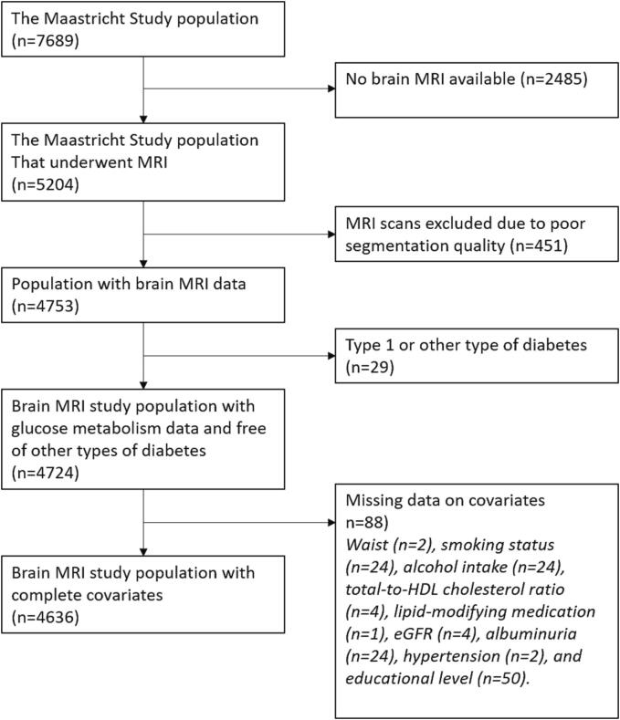
We used cross-sectional data from 4,724 participants (58.7 ± 8.5 years, 51.5% women) of The Maastricht Study, a population-based prospective cohort. Glucose metabolism status was assessed with an oral glucose tolerance test, and defined as type 2 diabetes (n = 869), prediabetes (n = 671), or normal glucose metabolism (n = 3184). We extracted 12 hippocampal subfield volumes per hemisphere with FreeSurfer v6.0 using T1w and FLAIR 3T MRI images. We used multiple linear regression and linear trend analysis, and adjusted for total intracranial volume, demographic, lifestyle, and cardiovascular risk factors.
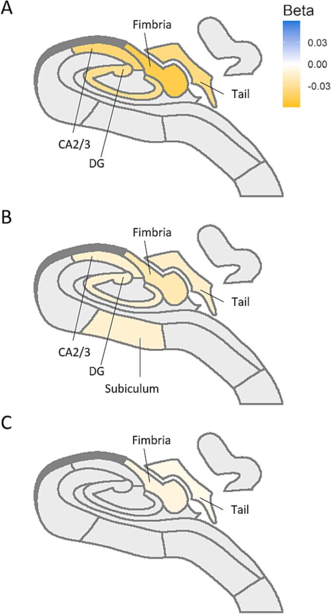
Type 2 diabetes was significantly associated with smaller volumes in the hippocampal subfield fimbria (standardized beta coefficient ± standard error (β ± SE) = -0.195 ± 0.04, p-value < 0.001), the hippocampus proper, i.e. Cornu Ammonis (CA) 1, CA2/3, CA4, dentate gyrus, subiculum and presubiculum (β ± SE < -0.105 ± 0.04, p-value < 0.006); as well as the hippocampal tail (β ± SE = -0.162 ± 0.04, p-value < 0.001). Prediabetes showed no significant associations. However, linear trend analysis indicated a dose-response relation from normal glucose metabolism, to prediabetes, to type 2 diabetes. Multiple continuous measures of hyperglycemia were associated with smaller volumes of the subfields fimbria (β ± SE < -0.010 ± 0.011, p-value < 0.001), dentate gyrus (β ± SE < -0.013 ± 0.010, p-value < 0.002), CA3 (β ± SE < -0.014 ± 0.011, p-value < 0.001), and tail (β ± SE < -0.006 ± 0.012, p-value < 0.003).
CONCLUSIONS/INTERPRETATION: Type 2 diabetes and measures of hyperglycemia are associated with hippocampal subfield atrophy, independently of lifestyle and cardiovascular risk factors. We found evidence for a dose-response relationship from normal glucose metabolism, to prediabetes, to type 2 diabetes. Prediabetes stages could give a window of opportunity for the early prevention of brain disease.
目的/假设:我们研究了前驱糖尿病、2 型糖尿病和持续的高血糖是否与海马体特定亚区的组织体积差异有关。
我们使用了 Maastricht 研究的横断面数据,该研究是一项基于人群的前瞻性队列研究,共有 4724 名参与者(58.7±8.5 岁,51.5%为女性)。葡萄糖代谢状态通过口服葡萄糖耐量试验进行评估,并定义为 2 型糖尿病(n=869)、前驱糖尿病(n=671)或正常葡萄糖代谢(n=3184)。我们使用 FreeSurfer v6.0 从 T1w 和 FLAIR 3T MRI 图像中提取每个半球的 12 个海马亚区体积。我们使用多元线性回归和线性趋势分析,并调整了总颅内体积、人口统计学、生活方式和心血管危险因素。
2 型糖尿病与海马体的穹窿(标准化β系数±标准误差(β±SE)=-0.195±0.04,p 值<0.001)、海马体本身(即 Cornu Ammonis [CA] 1、CA2/3、CA4、齿状回、下托和前下托)的体积缩小显著相关(β±SE <-0.105±0.04,p 值<0.006);以及海马体尾部(β±SE=-0.162±0.04,p 值<0.001)。前驱糖尿病没有明显的相关性。然而,线性趋势分析表明,从正常葡萄糖代谢到前驱糖尿病再到 2 型糖尿病,存在剂量反应关系。多个连续的高血糖指标与穹窿(β±SE <-0.010±0.011,p 值<0.001)、齿状回(β±SE <-0.013±0.010,p 值<0.002)、CA3(β±SE <-0.014±0.011,p 值<0.001)和尾部(β±SE <-0.006±0.012,p 值<0.003)的体积缩小有关。
结论/解释:2 型糖尿病和高血糖指标与海马体亚区萎缩有关,独立于生活方式和心血管危险因素。我们发现,从正常葡萄糖代谢到前驱糖尿病再到 2 型糖尿病,存在剂量反应关系。前驱糖尿病阶段可能为预防脑部疾病提供了一个早期机会。