• 文献检索
  • 文档翻译
  • 深度研究
  • 学术资讯
  • Suppr Zotero 插件Zotero 插件
  • 邀请有礼
  • 套餐&价格
  • 历史记录
应用&插件
Suppr Zotero 插件Zotero 插件浏览器插件Mac 客户端Windows 客户端微信小程序
定价
高级版会员购买积分包购买API积分包
服务
文献检索文档翻译深度研究API 文档MCP 服务
关于我们
关于 Suppr公司介绍联系我们用户协议隐私条款
关注我们

Suppr 超能文献

核心技术专利:CN118964589B侵权必究
粤ICP备2023148730 号-1Suppr @ 2026

文献检索

告别复杂PubMed语法,用中文像聊天一样搜索,搜遍4000万医学文献。AI智能推荐,让科研检索更轻松。

立即免费搜索

文件翻译

保留排版,准确专业,支持PDF/Word/PPT等文件格式,支持 12+语言互译。

免费翻译文档

深度研究

AI帮你快速写综述,25分钟生成高质量综述,智能提取关键信息,辅助科研写作。

立即免费体验

基于钆塞酸二钠增强 MRI 的非酒精性脂肪性肝病患者的组织学、影像学和人工智能特征的相关性:一项概念验证研究。

Correlation of histologic, imaging, and artificial intelligence features in NAFLD patients, derived from Gd-EOB-DTPA-enhanced MRI: a proof-of-concept study.

机构信息

Department of Biomedical Imaging and Image-Guided Therapy, Medical University of Vienna, Vienna, Austria.

Computational Imaging Research Lab, Department of Biomedical Imaging and Image-Guided Therapy, Medical University of Vienna, Vienna, Austria.

出版信息

Eur Radiol. 2023 Nov;33(11):7729-7743. doi: 10.1007/s00330-023-09735-5. Epub 2023 Jun 26.

DOI:10.1007/s00330-023-09735-5
PMID:37358613
原文链接:https://pmc.ncbi.nlm.nih.gov/articles/PMC10598123/
Abstract

OBJECTIVE

To compare unsupervised deep clustering (UDC) to fat fraction (FF) and relative liver enhancement (RLE) on Gd-EOB-DTPA-enhanced MRI to distinguish simple steatosis from non-alcoholic steatohepatitis (NASH), using histology as the gold standard.

MATERIALS AND METHODS

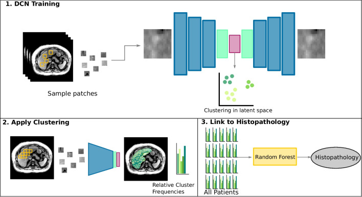
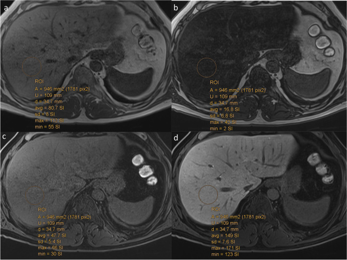
A derivation group of 46 non-alcoholic fatty liver disease (NAFLD) patients underwent 3-T MRI. Histology assessed steatosis, inflammation, ballooning, and fibrosis. UDC was trained to group different texture patterns from MR data into 10 distinct clusters per sequence on unenhanced T1- and Gd-EOB-DTPA-enhanced T1-weighted hepatobiliary phase (T1-Gd-EOB-DTPA-HBP), then on T1 in- and opposed-phase images. RLE and FF were quantified on identical sequences. Differences of these parameters between NASH and simple steatosis were evaluated with χ- and t-tests, respectively. Linear regression and Random Forest classifier were performed to identify associations between histological NAFLD features, RLE, FF, and UDC patterns, and then determine predictors able to distinguish simple steatosis from NASH. ROC curves assessed diagnostic performance of UDC, RLE, and FF. Finally, we tested these parameters on 30 validation cohorts.

RESULTS

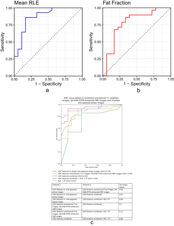
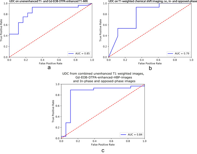
For the derivation group, UDC-derived features from unenhanced and T1-Gd-EOB-DTPA-HBP, plus from T1 in- and opposed-phase, distinguished NASH from simple steatosis (p ≤ 0.001 and p = 0.02, respectively) with 85% and 80% accuracy, respectively, while RLE and FF distinguished NASH from simple steatosis (p ≤ 0.001 and p = 0.004, respectively), with 83% and 78% accuracy, respectively. On multivariate regression analysis, RLE and FF correlated only with fibrosis (p = 0.040) and steatosis (p ≤ 0.001), respectively. Conversely, UDC features, using Random Forest classifier predictors, correlated with all histologic NAFLD components. The validation group confirmed these results for both approaches.

CONCLUSION

UDC, RLE, and FF could independently separate NASH from simple steatosis. UDC may predict all histologic NAFLD components.

CLINICAL RELEVANCE STATEMENT

Using gadoxetic acid-enhanced MR, fat fraction (FF > 5%) can diagnose NAFLD, and relative liver enhancement can distinguish NASH from simple steatosis. Adding AI may let us non-invasively estimate the histologic components, i.e., fat, ballooning, inflammation, and fibrosis, the latter the main prognosticator.

KEY POINTS

• Unsupervised deep clustering (UDC) and MR-based parameters (FF and RLE) could independently distinguish simple steatosis from NASH in the derivation group. • On multivariate analysis, RLE could predict only fibrosis, and FF could predict only steatosis; however, UDC could predict all histologic NAFLD components in the derivation group. • The validation cohort confirmed the findings for the derivation group.

摘要

目的

使用钆塞酸二钠增强 MRI 的无监督深度聚类(UDC)与脂肪分数(FF)和相对肝增强(RLE)比较,以组织学为金标准,区分单纯性脂肪变性与非酒精性脂肪性肝炎(NASH)。

材料与方法

对 46 名非酒精性脂肪性肝病(NAFLD)患者进行 3-T MRI 检查。组织学评估脂肪变性、炎症、气球样变和纤维化。UDC 用于对未增强 T1 加权和 Gd-EOB-DTPA 增强 T1 期肝胆期(T1-Gd-EOB-DTPA-HBP)的 MR 数据中的不同纹理模式进行分组,然后对 T1 同相位和反相位图像进行分组。在相同的序列上量化 RLE 和 FF。使用 χ 和 t 检验分别评估这些参数在 NASH 和单纯性脂肪变性之间的差异。进行线性回归和随机森林分类器分析,以确定组织学 NAFLD 特征、RLE、FF 和 UDC 模式之间的关联,然后确定能够区分单纯性脂肪变性和 NASH 的预测因子。ROC 曲线评估 UDC、RLE 和 FF 的诊断性能。最后,我们在 30 个验证队列中测试了这些参数。

结果

在推导组中,来自未增强和 T1-Gd-EOB-DTPA-HBP 的 UDC 衍生特征,加上 T1 同相位和反相位的 UDC 衍生特征,可区分 NASH 与单纯性脂肪变性(p≤0.001 和 p=0.02),准确性分别为 85%和 80%,而 RLE 和 FF 可区分 NASH 与单纯性脂肪变性(p≤0.001 和 p=0.004),准确性分别为 83%和 78%。多元回归分析显示,RLE 和 FF 仅与纤维化(p=0.040)和脂肪变性(p≤0.001)相关。相反,UDC 特征,使用随机森林分类器预测因子,与所有组织学 NAFLD 成分相关。验证组对这两种方法均证实了这些结果。

结论

UDC、RLE 和 FF 可独立区分 NASH 与单纯性脂肪变性。UDC 可能预测所有组织学 NAFLD 成分。

临床相关性

使用钆塞酸增强 MRI,脂肪分数(FF>5%)可诊断 NAFLD,相对肝增强可区分 NASH 与单纯性脂肪变性。添加 AI 可能使我们能够无创性估计组织学成分,即脂肪、气球样变、炎症和纤维化,后者是主要的预后因素。

https://cdn.ncbi.nlm.nih.gov/pmc/blobs/6de6/10598123/9e568ae1d5a5/330_2023_9735_Fig4_HTML.jpg
https://cdn.ncbi.nlm.nih.gov/pmc/blobs/6de6/10598123/18cac68a7a45/330_2023_9735_Fig1_HTML.jpg
https://cdn.ncbi.nlm.nih.gov/pmc/blobs/6de6/10598123/34cf32e9de70/330_2023_9735_Fig2_HTML.jpg
https://cdn.ncbi.nlm.nih.gov/pmc/blobs/6de6/10598123/dd9e7f75e6dd/330_2023_9735_Fig3_HTML.jpg
https://cdn.ncbi.nlm.nih.gov/pmc/blobs/6de6/10598123/9e568ae1d5a5/330_2023_9735_Fig4_HTML.jpg

相似文献

1
Correlation of histologic, imaging, and artificial intelligence features in NAFLD patients, derived from Gd-EOB-DTPA-enhanced MRI: a proof-of-concept study.基于钆塞酸二钠增强 MRI 的非酒精性脂肪性肝病患者的组织学、影像学和人工智能特征的相关性:一项概念验证研究。
Eur Radiol. 2023 Nov;33(11):7729-7743. doi: 10.1007/s00330-023-09735-5. Epub 2023 Jun 26.
2
Gd-EOB-DTPA-enhanced-MR imaging in the inflammation stage of nonalcoholic steatohepatitis (NASH) in mice.钆塞酸二钠增强磁共振成像在小鼠非酒精性脂肪性肝炎(NASH)炎症期的应用
Magn Reson Imaging. 2016 Jul;34(6):724-729. doi: 10.1016/j.mri.2016.03.009. Epub 2016 Mar 12.
3
Usefulness of T1 mapping on Gd-EOB-DTPA-enhanced MR imaging in assessment of non-alcoholic fatty liver disease.钆塞酸二钠增强磁共振成像 T1 mapping 在非酒精性脂肪性肝病评估中的作用。
Eur Radiol. 2014 Apr;24(4):959-66. doi: 10.1007/s00330-014-3096-y. Epub 2014 Jan 26.
4
Non-contrast T1ρ dispersion versus Gd-EOB-DTPA-enhanced T1mapping for the risk stratification of non-alcoholic fatty liver disease in rabbit models.非对比T1ρ弥散与钆塞酸二钠增强T1成像用于兔模型中非酒精性脂肪性肝病风险分层的研究
Magn Reson Imaging. 2024 Apr;107:130-137. doi: 10.1016/j.mri.2024.01.013. Epub 2024 Jan 24.
5
Usefulness of Gd-EOB-DTPA-enhanced MR imaging in the evaluation of simple steatosis and nonalcoholic steatohepatitis.钆塞酸二钠增强 MRI 在单纯性脂肪变性和非酒精性脂肪性肝炎评估中的作用。
J Magn Reson Imaging. 2013 May;37(5):1137-43. doi: 10.1002/jmri.23921. Epub 2012 Nov 21.
6
Gd-EOB-DTPA-enhanced T1ρ imaging vs diffusion metrics for assessment liver inflammation and early stage fibrosis of nonalcoholic steatohepatitis in rabbits.钆塞酸二钠增强T1ρ成像与扩散指标用于评估兔非酒精性脂肪性肝炎的肝脏炎症和早期纤维化
Magn Reson Imaging. 2018 May;48:34-41. doi: 10.1016/j.mri.2017.12.017. Epub 2017 Dec 24.
7
Evaluation of hepatic function using dynamic contrast-enhanced magnetic resonance imaging in melanocortin 4 receptor-deficient mice as a model of nonalcoholic steatohepatitis.使用动态对比增强磁共振成像评估黑素皮质素4受体缺陷小鼠作为非酒精性脂肪性肝炎模型的肝功能。
Magn Reson Imaging. 2019 Apr;57:210-217. doi: 10.1016/j.mri.2018.11.013. Epub 2018 Nov 19.
8
The natural history of streptozotocin-stimulated non-alcoholic steatohepatitis mice followed by Gd-EOB-DTPA-enhanced MRI: Comparison with simple steatosis mice.链脲佐菌素诱导的非酒精性脂肪性肝炎小鼠经钆塞酸二钠增强磁共振成像后的自然病程:与单纯性脂肪肝小鼠的比较
Magn Reson Imaging. 2017 May;38:123-128. doi: 10.1016/j.mri.2016.12.027. Epub 2017 Jan 3.
9
Multiparametric MR Index for the Diagnosis of Non-Alcoholic Steatohepatitis in Patients with Non-Alcoholic Fatty Liver Disease.多参数磁共振指数在非酒精性脂肪性肝病患者非酒精性肝炎诊断中的应用。
Sci Rep. 2020 Feb 14;10(1):2671. doi: 10.1038/s41598-020-59601-3.
10
Gadolinium-ethoxybenzyl-diethylenetriamine penta-acetic acid-enhanced magnetic resonance imaging for evaluating fibrosis regression in chronic hepatitis C patients after direct-acting antiviral.钆特醇乙氧苯甲基二乙三胺五乙酸增强磁共振成像用于评估直接作用抗病毒治疗后慢性丙型肝炎患者纤维化的逆转。
World J Gastroenterol. 2022 May 28;28(20):2214-2226. doi: 10.3748/wjg.v28.i20.2214.

本文引用的文献

1
Mechanisms and disease consequences of nonalcoholic fatty liver disease.非酒精性脂肪性肝病的机制及疾病后果
Cell. 2021 May 13;184(10):2537-2564. doi: 10.1016/j.cell.2021.04.015.
2
High-Throughput, Machine Learning-Based Quantification of Steatosis, Inflammation, Ballooning, and Fibrosis in Biopsies From Patients With Nonalcoholic Fatty Liver Disease.高通量、基于机器学习的非酒精性脂肪性肝病患者肝活检组织中脂肪变性、炎症、气球样变、纤维化的定量分析。
Clin Gastroenterol Hepatol. 2020 Aug;18(9):2081-2090.e9. doi: 10.1016/j.cgh.2019.12.025. Epub 2019 Dec 27.
3
Noninvasive evaluation of nonalcoholic fatty liver disease: Current evidence and practice.
非酒精性脂肪性肝病的无创评估:当前证据与实践
World J Gastroenterol. 2019 Mar 21;25(11):1307-1326. doi: 10.3748/wjg.v25.i11.1307.
4
Noninvasive, Quantitative Assessment of Liver Fat by MRI-PDFF as an Endpoint in NASH Trials.MRI-PDFF 无创定量评估肝脂肪作为 NASH 试验的终点。
Hepatology. 2018 Aug;68(2):763-772. doi: 10.1002/hep.29797.
5
MR elastography is effective for the non-invasive evaluation of fibrosis and necroinflammatory activity in patients with nonalcoholic fatty liver disease.磁共振弹性成像可有效用于非酒精性脂肪性肝病患者肝纤维化和坏死性炎症活动的无创评估。
Eur J Radiol. 2018 Jan;98:82-89. doi: 10.1016/j.ejrad.2017.11.003. Epub 2017 Nov 11.
6
Non-invasive assessment of non-alcoholic fatty liver disease: Clinical prediction rules and blood-based biomarkers.非酒精性脂肪性肝病的无创评估:临床预测规则和基于血液的生物标志物。
J Hepatol. 2018 Feb;68(2):305-315. doi: 10.1016/j.jhep.2017.11.013. Epub 2017 Dec 2.
7
Global burden of NAFLD and NASH: trends, predictions, risk factors and prevention.非酒精性脂肪性肝病和非酒精性脂肪性肝炎的全球负担:趋势、预测、危险因素和预防。
Nat Rev Gastroenterol Hepatol. 2018 Jan;15(1):11-20. doi: 10.1038/nrgastro.2017.109. Epub 2017 Sep 20.
8
MR Spectroscopy-derived Proton Density Fat Fraction Is Superior to Controlled Attenuation Parameter for Detecting and Grading Hepatic Steatosis.磁共振波谱质子密度脂肪分数检测与分级肝脂肪变优于衰减参数控制。
Radiology. 2018 Feb;286(2):547-556. doi: 10.1148/radiol.2017162931. Epub 2017 Sep 15.
9
Linearity, Bias, and Precision of Hepatic Proton Density Fat Fraction Measurements by Using MR Imaging: A Meta-Analysis.使用磁共振成像测量肝脏质子密度脂肪分数的线性、偏差和精密度:一项荟萃分析
Radiology. 2018 Feb;286(2):486-498. doi: 10.1148/radiol.2017170550. Epub 2017 Sep 11.
10
The diagnosis and management of nonalcoholic fatty liver disease: Practice guidance from the American Association for the Study of Liver Diseases.非酒精性脂肪性肝病的诊断与管理:美国肝病研究协会的实践指南
Hepatology. 2018 Jan;67(1):328-357. doi: 10.1002/hep.29367. Epub 2017 Sep 29.