Guo Jiaqi, Zhang Dan, Gong Yan, Liu Jiang, Zhang Jiong, Zhao Yitian
Cixi Institute of Biomedical Engineering, Ningbo Institute of Materials Technology and Engineering, Chinese Academy of Sciences, Ningbo, China.
University of Chinese Academy of Sciences, Beijing, China.
Front Neurosci. 2023 Jun 9;17:1194661. doi: 10.3389/fnins.2023.1194661. eCollection 2023.
Neuromyelitis optica spectrum disorders (NMOSD) are autoimmune central nervous system diseases characterized by the immune system's abnormal attack on glial cells and neurons. Optic neuritis (ON) is one of the indicators of NMOSD, often starting unilaterally and potentially affecting both eyes later in the disease progression, leading to visual impairment. Optical coherence tomography angiography (OCTA) has the potential to aid in the early diagnosis of NMOSD by examining ophthalmic imaging and may offer a window for disease prevention.
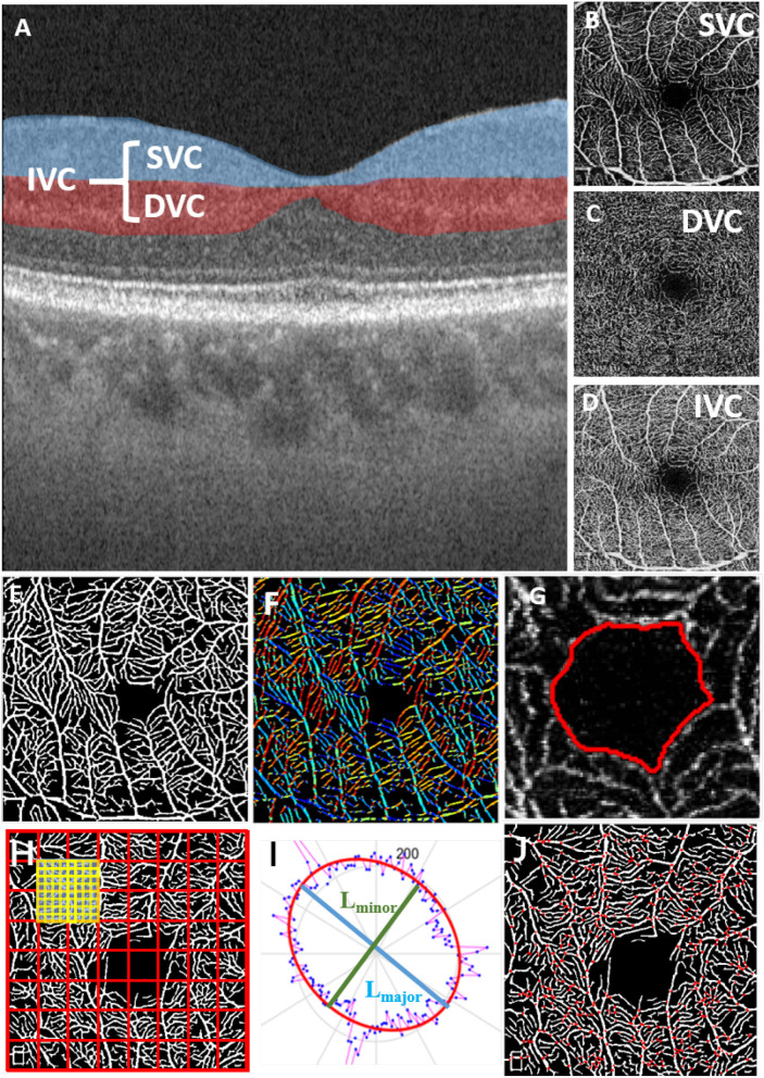
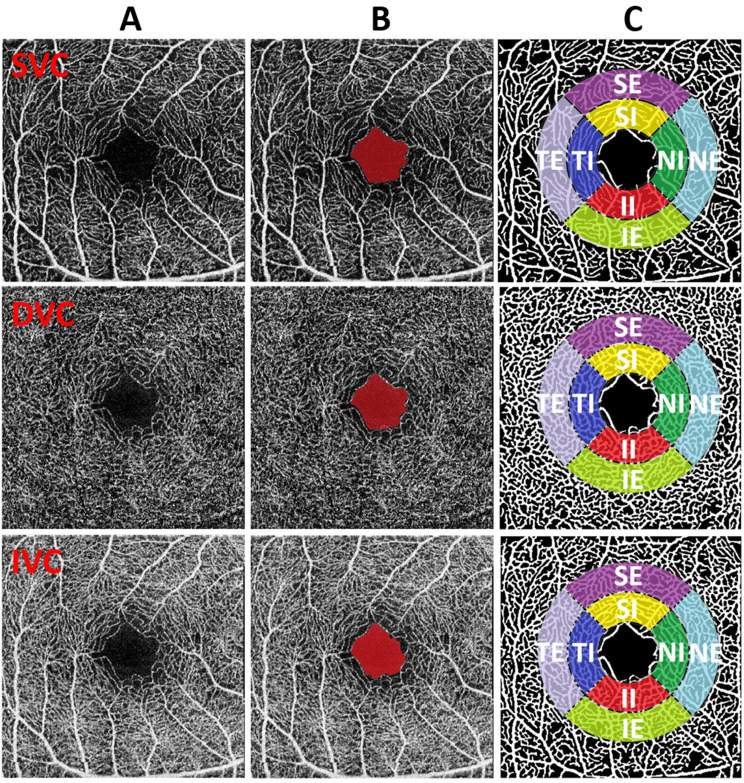
In this study, we collected OCTA images from 22 NMOSD patients (44 images) and 25 healthy individuals (50 images) to investigate retinal microvascular changes in NMOSD. We employed effective retinal microvascular segmentation and foveal avascular zone (FAZ) segmentation techniques to extract key OCTA structures for biomarker analysis. A total of 12 microvascular features were extracted using specifically designed methods based on the segmentation results. The OCTA images of NMOSD patients were classified into two groups: optic neuritis (ON) and non-optic neuritis (non-ON). Each group was compared separately with a healthy control (HC) group.
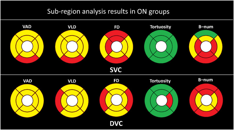
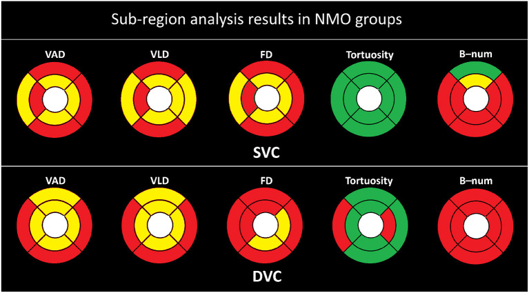
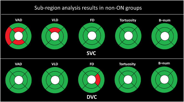
Statistical analysis revealed that the non-ON group displayed shape changes in the deep layer of the retina, specifically in the FAZ. However, there were no significant microvascular differences between the non-ON group and the HC group. In contrast, the ON group exhibited microvascular degeneration in both superficial and deep retinal layers. Sub-regional analysis revealed that pathological variations predominantly occurred on the side affected by ON, particularly within the internal ring near the FAZ.
The findings of this study highlight the potential of OCTA in evaluating retinal microvascular changes associated with NMOSD. The shape alterations observed in the FAZ of the non-ON group suggest localized vascular abnormalities. In the ON group, microvascular degeneration in both superficial and deep retinal layers indicates more extensive vascular damage. Sub-regional analysis further emphasizes the impact of optic neuritis on pathological variations, particularly near the FAZ's internal ring.
This study provides insights into the retinal microvascular changes associated with NMOSD using OCTA imaging. The identified biomarkers and observed alterations may contribute to the early diagnosis and monitoring of NMOSD, potentially offering a time window for intervention and prevention of disease progression.
视神经脊髓炎谱系障碍(NMOSD)是自身免疫性中枢神经系统疾病,其特征是免疫系统对神经胶质细胞和神经元进行异常攻击。视神经炎(ON)是NMOSD的指标之一,通常单眼起病,在疾病进展后期可能累及双眼,导致视力损害。光学相干断层扫描血管造影(OCTA)有潜力通过检查眼科成像辅助NMOSD的早期诊断,并可能为疾病预防提供一个窗口。
在本研究中,我们收集了22例NMOSD患者的OCTA图像(44幅图像)和25名健康个体的OCTA图像(50幅图像),以研究NMOSD患者的视网膜微血管变化。我们采用有效的视网膜微血管分割和黄斑无血管区(FAZ)分割技术来提取关键的OCTA结构用于生物标志物分析。基于分割结果,使用专门设计的方法共提取了12个微血管特征。将NMOSD患者的OCTA图像分为两组:视神经炎(ON)组和非视神经炎(非ON)组。每组分别与健康对照组(HC)进行比较。
统计分析显示,非ON组在视网膜深层,特别是在FAZ处显示出形状变化。然而,非ON组与HC组之间微血管差异不显著。相比之下,ON组在视网膜浅层和深层均表现出微血管变性。亚区域分析显示,病理变化主要发生在ON受累侧,特别是在FAZ附近的内环内。
本研究结果突出了OCTA在评估与NMOSD相关的视网膜微血管变化方面的潜力。在非ON组FAZ中观察到的形状改变提示局部血管异常。在ON组中,视网膜浅层和深层的微血管变性表明血管损伤更广泛。亚区域分析进一步强调了视神经炎对病理变化的影响,特别是在FAZ内环附近。
本研究使用OCTA成像深入了解了与NMOSD相关的视网膜微血管变化。所确定的生物标志物和观察到的改变可能有助于NMOSD的早期诊断和监测, potentially offering a time window for intervention and prevention of disease progression.(原文最后一句英文表述有误,正确表述应该是“potentially offering a time window for intervention and prevention of disease progression”,译文:可能为干预和预防疾病进展提供一个时间窗口 ) 从而可能为干预和预防疾病进展提供一个时间窗口。