Experimental and Clinical Research Center, Max Delbrueck Center for Molecular Medicine and Charité - Universitätsmedizin Berlin, corporate member of Freie Universität Berlin, Humboldt-Universität zu Berlin, and Berlin Institute of Health, Berlin, Germany; NeuroCure Clinical Research Center, Charité - Universitätsmedizin Berlin, corporate member of Freie Universität Berlin, Humboldt-Universität zu Berlin, and Berlin Institute of Health, Berlin, Germany; Neurologic Clinic and Policlinic, Department of Medicine, Clinical Research and Biomedicine University Hospital Basel, University of Basel, Basel, Switzerland.
Experimental and Clinical Research Center, Max Delbrueck Center for Molecular Medicine and Charité - Universitätsmedizin Berlin, corporate member of Freie Universität Berlin, Humboldt-Universität zu Berlin, and Berlin Institute of Health, Berlin, Germany; NeuroCure Clinical Research Center, Charité - Universitätsmedizin Berlin, corporate member of Freie Universität Berlin, Humboldt-Universität zu Berlin, and Berlin Institute of Health, Berlin, Germany; Department of Neurology, University of California San Francisco, CA, USA.
Neuroimage Clin. 2021;30:102608. doi: 10.1016/j.nicl.2021.102608. Epub 2021 Mar 4.
Lateral geniculate nucleus (LGN) volume is reduced after optic neuritis (ON) in neuromyelitis optica spectrum disorders (NMOSD). We aimed at a longitudinal assessment of LGN volume in NMOSD.
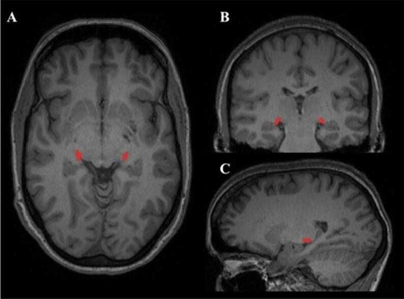
Twenty-nine patients with aquaporin 4-IgG seropositive NMOSD (age: 47.8 ± 14.6 years (y), female: n = 27, history of ON (NMO-ON): n = 17, median time since ON: 3[1.2-12.1]y) and 18 healthy controls (HC; age: 39.3 ± 15.8y; female: n = 13) were included. Median follow-up was 4.1[1.1-4.7]y for patients and 1.7[0.9-3.2]y for HC. LGN volume was measured using a multi-atlas-based approach of automated segmentation on 3 Tesla magnetic resonance images. Retinal optical coherence tomography and probabilistic tractography of the optic radiations (OR) were also performed.
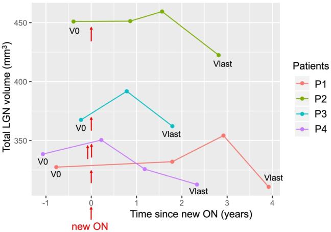
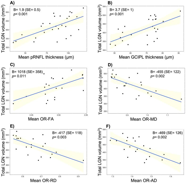
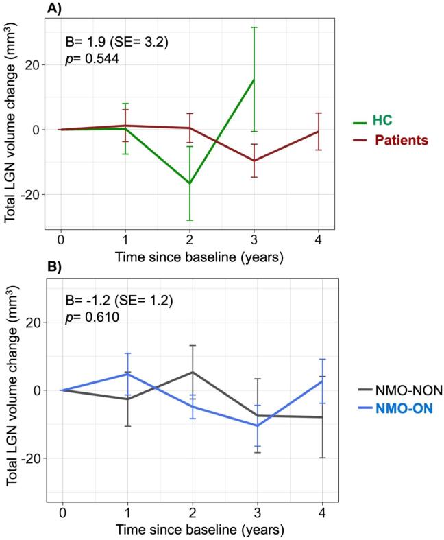
At baseline, NMO-ON patients had lower LGN volumes (395.4 ± 48.9 mm) than patients without ON (NMO-NON: 450.7 ± 55.6 mm; p = 0.049) and HC (444.5 ± 61.5 mm, p = 0.025). LGN volume was associated with retinal neuroaxonal loss and microstructural OR damage. Longitudinally, there was no change in LGN volumes in the absence of ON, neither in all patients (B = -0.6, SE = 1.4, p = 0.670), nor in NMO-ON (B = -0.8, SE = 1.6, p = 0.617) and NMO-NON (B = 1.7, SE = 3.5, p = 0.650). However, in four patients with new ON during follow-up, LGN volume was reduced at last visit (median time since ON: 2.6 [1.8-3.9]y) compared to the measurement before ON (352 ± 52.7 vs. 371.1 ± 55.9 mm; t = -3.6, p = 0.036).
Although LGN volume is reduced after ON in NMOSD, this volume loss is not progressive over longer follow-up or independent of ON. Thus, our findings -at least in this relatively small cohort- do not support occult neurodegeneration of the afferent visual pathway in NMOSD.
在视神经脊髓炎(NMOSD)谱系疾病中,外侧膝状体(LGN)体积在视神经炎(ON)后减少。我们旨在对 NMOSD 中的 LGN 体积进行纵向评估。
纳入 29 例水通道蛋白 4-IgG 阳性 NMOSD 患者(年龄:47.8±14.6 岁(y),女性:n=27,ON 病史(NMO-ON):n=17,ON 后中位时间:3[1.2-12.1]y)和 18 名健康对照者(HC;年龄:39.3±15.8y;女性:n=13)。患者的中位随访时间为 4.1[1.1-4.7]y,HC 为 1.7[0.9-3.2]y。使用基于多图谱的自动分割方法在 3T 磁共振图像上测量 LGN 体积。还进行了视网膜光学相干断层扫描和视辐射(OR)的概率追踪。
基线时,NMO-ON 患者的 LGN 体积(395.4±48.9mm)低于无 ON(NMO-NON:450.7±55.6mm;p=0.049)和 HC(444.5±61.5mm,p=0.025)。LGN 体积与视网膜神经轴突丢失和 OR 的微观结构损伤有关。纵向来看,在没有 ON 的情况下,LGN 体积没有变化,无论是在所有患者中(B=-0.6,SE=1.4,p=0.670),还是在 NMO-ON 中(B=-0.8,SE=1.6,p=0.617)和 NMO-NON 中(B=1.7,SE=3.5,p=0.650)。然而,在随访期间出现新 ON 的 4 名患者中,与 ON 前的测量值相比,最后一次就诊时的 LGN 体积(ON 后中位时间:2.6[1.8-3.9]y)减少了(352±52.7 vs. 371.1±55.9mm;t=-3.6,p=0.036)。
尽管 NMOSD 中的 ON 后 LGN 体积减少,但在较长时间的随访中,这种体积损失并不是进行性的,也与 ON 无关。因此,我们的发现——至少在这个相对较小的队列中——不支持 NMOSD 中传入视觉通路的隐匿性神经退行性变。