Center for Molecular Biophysics UPR4301 CNRS, Rue Charles Sadron, 45071 Orléans Cedex 2, France.
Oxid Med Cell Longev. 2023 Jun 17;2023:6829931. doi: 10.1155/2023/6829931. eCollection 2023.
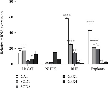
Keratinocytes prevent skin photoaging by ensuring the defence against oxidative stress, an excessive production of reactive oxygen species (ROS). They are localized within the epidermis where the oxygen level (1-3% O), named physioxia, is low compared to other organs. Oxygen is essential for life but also generates ROS. Most of the studies on keratinocyte antioxidant capacities are performed under atmospheric oxygen, named normoxia, which is very far from the physiological microenvironment, thus submitting cells to an overoxygenation. The present study is aimed at investigating the antioxidant status of keratinocyte grown under physioxia in both 2D and 3D models. First, we show that the basal antioxidant profiles of keratinocytes display important differences when comparing the HaCaT cell line, primary keratinocytes (NHEK), reconstructed epidermis (RHE), and skin explants. Physioxia was shown to promote a strong proliferation of keratinocytes in monolayers and in RHE, resulting in a thinner epidermis likely due to a slowdown in cell differentiation. Interestingly, cells in physioxia exhibited a lower ROS production upon stress, suggesting a better protection against oxidative stress. To understand this effect, we studied the antioxidant enzymes and reported a lower or equivalent level of mRNA for all enzymes in physioxia conditions compared to normoxia, but a higher activity for catalase and superoxide dismutases, whatever the culture model. The unchanged catalase amount, in NHEK and RHE, suggests an overactivation of the enzyme in physioxia, whereas the higher amount of SOD2 can explain the strong activity. Taken together, our results demonstrate the role of oxygen in the regulation of the antioxidant defences in keratinocytes, topic of particular importance for studying skin aging. Additionally, the present work points out the interest of the choice of both the keratinocyte culture model and the oxygen level to be as close as possible to the skin.
角质形成细胞通过确保对氧化应激(活性氧(ROS)的过度产生)的防御来防止皮肤光老化。它们位于表皮内,其中氧水平(1-3% O),称为低氧,与其他器官相比很低。氧气是生命所必需的,但也会产生 ROS。大多数关于角质形成细胞抗氧化能力的研究都是在大气氧(称为常氧)下进行的,这与生理微环境相差甚远,从而使细胞过度氧化。本研究旨在研究在 2D 和 3D 模型中低氧培养的角质形成细胞的抗氧化状态。首先,我们表明,比较 HaCaT 细胞系、原代角质形成细胞(NHEK)、重建表皮(RHE)和皮肤外植体时,角质形成细胞的基础抗氧化谱存在重要差异。低氧促进了单层和 RHE 中角质形成细胞的强烈增殖,导致表皮变薄,可能是由于细胞分化减慢。有趣的是,低氧细胞在应激时产生的 ROS 产生较少,表明对氧化应激的保护更好。为了理解这种影响,我们研究了抗氧化酶,并报告了在低氧条件下与常氧相比,所有酶的 mRNA 水平较低或相等,但过氧化氢酶和超氧化物歧化酶的活性更高,无论培养模型如何。在 NHEK 和 RHE 中不变的过氧化氢酶量表明该酶在低氧下过度激活,而 SOD2 的高含量可以解释其高活性。总之,我们的结果表明了氧在调节角质形成细胞抗氧化防御中的作用,这对于研究皮肤衰老具有特别重要的意义。此外,本工作指出了选择角质形成细胞培养模型和氧水平尽可能接近皮肤的重要性。