Peterson Michael W, Jain Ratnali, Hildebrandt Kurt, Carson William Keith, Fayed Mohamed A
Fresno Department of Medicine, University of California (San Francisco), San Francisco, CA 93701, USA.
UCSF Fresno/Community Medical Centers' Multidisciplinary Lung Nodule Clinic, Fresno, CA 93701, USA.
J Fungi (Basel). 2023 Jun 1;9(6):641. doi: 10.3390/jof9060641.
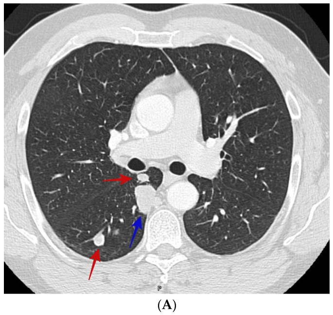
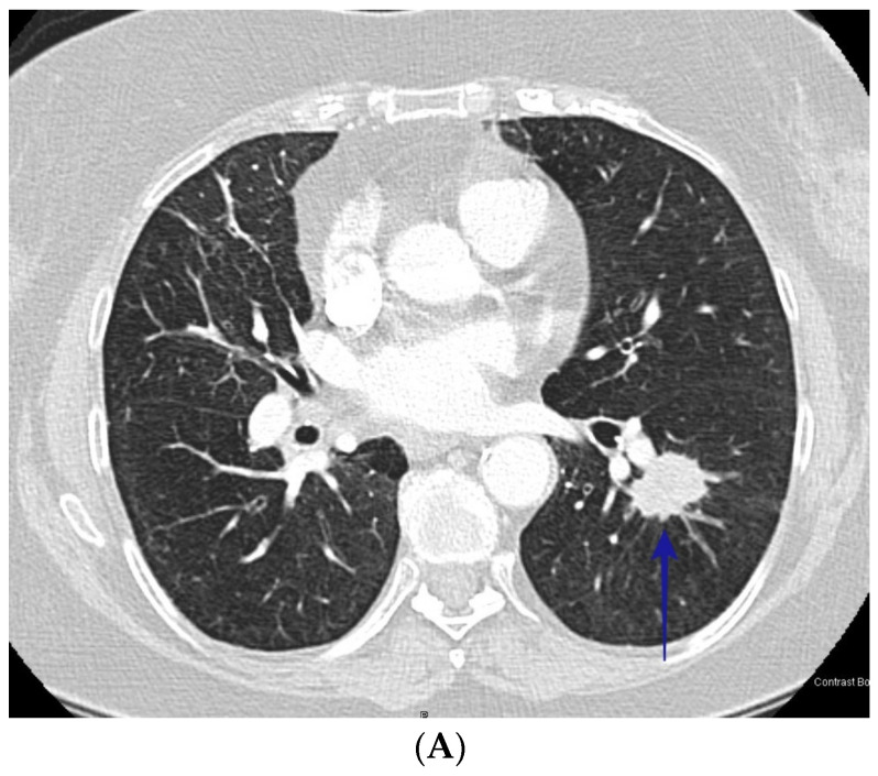
Coccidioidomycosis (cocci) is an endemic fungal disease that can cause asymptomatic or post-symptomatic lung nodules which are visible on chest CT scanning. Lung nodules are common and can represent early lung cancer. Differentiating lung nodules due to cocci from those due to lung cancer can be difficult and lead to invasive and expensive evaluations.
We identified 302 patients with biopsy-proven cocci or bronchogenic carcinoma seen in our multidisciplinary nodule clinic. Two experienced radiologists who were blinded to the diagnosis read the chest CT scans and identified radiographic characteristics to determine their utility in differentiating lung cancer nodules from those due to cocci.
Using univariate analysis, we identified several radiographic findings that differed between lung cancer and cocci infection. We then entered these variables along with age and gender into a multivariate model and found that age, nodule diameter, nodule cavitation, presence of satellite nodules and radiographic presence of chronic lung disease differed significantly between the two diagnoses. Three findings, cavitary nodules, satellite nodules and chronic lung disease, have sufficient discrimination to potentially be useful in clinical decision-making.
Careful evaluation of the three obtained radiographic findings can significantly improve our ability to differentiate benign coccidioidomycosis infection from lung cancer in an endemic region for the fungal disease. Using these data may significantly reduce the cost and risk associated with distinguishing the cause of lung nodules in these patients by preventing unnecessary invasive studies.
球孢子菌病是一种地方性真菌病,可导致无症状或症状后肺结节,在胸部CT扫描中可见。肺结节很常见,可能代表早期肺癌。区分由球孢子菌引起的肺结节和由肺癌引起的肺结节可能很困难,并导致侵入性和昂贵的评估。
我们在多学科结节门诊中确定了302例经活检证实患有球孢子菌病或支气管源性癌的患者。两位对诊断不知情的经验丰富的放射科医生阅读胸部CT扫描,并确定影像学特征,以确定其在区分肺癌结节和球孢子菌病结节方面的效用。
通过单变量分析,我们确定了肺癌和球孢子菌感染之间存在差异的几个影像学表现。然后,我们将这些变量以及年龄和性别纳入多变量模型,发现年龄、结节直径、结节空洞、卫星结节的存在以及慢性肺病的影像学表现在两种诊断之间存在显著差异。三个表现,即空洞结节、卫星结节和慢性肺病,具有足够的鉴别力,可能在临床决策中有用。
仔细评估获得的这三个影像学表现可显著提高我们在真菌病流行地区区分良性球孢子菌病感染和肺癌的能力。利用这些数据可能会通过避免不必要的侵入性研究,显著降低与区分这些患者肺结节病因相关的成本和风险。