Department of Pulmonary and Critical Care Medicine, Lung Cancer Center, West China Hospital, Sichuan University, Precision Medicine Key Laboratory of Sichuan Province, Chengdu 610041, China
Biomolecules. 2023 Jun 15;13(6):991. doi: 10.3390/biom13060991.
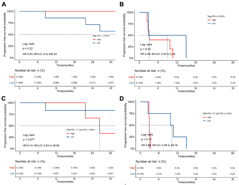
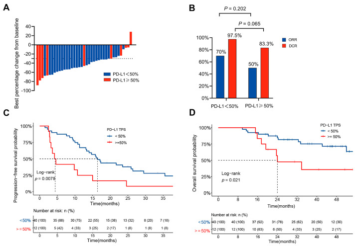
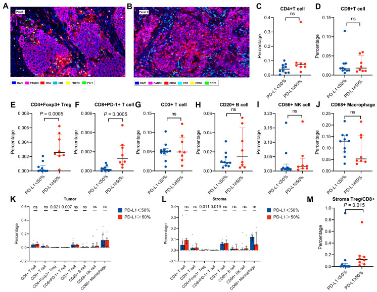
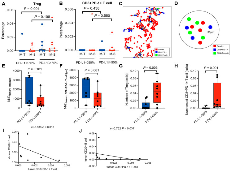
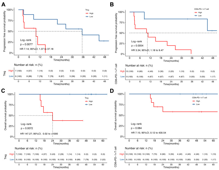
High tumour programmed cell death-ligand 1 (PD-L1) expression is associated with poor progression-free survival (PFS) after tyrosine kinase inhibitor (TKI) therapy in -rearranged non-small cell lung cancer (NSCLC). However, the characteristics of the tumour microenvironment (TME) and their prognostic values in -rearranged NSCLC are unknown. Here, we collected tumour tissues from pretreated -rearranged NSCLC patients, immunohistochemical staining was used to assess PD-L1 expression, and tumour-infiltrating immune cells were determined via multiplex immunofluorescence staining (mIF). Our data showed that the median values of PFS for the high PD-L1 group and low PD-L1 group who received ALK-TKI treatment were 4.4 and 16.4 months, respectively ( = 0.008). The median overall survival (OS) of the two groups was 24.0 months and not reached, respectively ( = 0.021). Via univariate and multivariate analyses, a high PD-L1 expression and a worse ECOG PS were determined to be independent prognostic factors of OS (HR = 3.35, 95% CI: 1.23-9.11, = 0.018; HR = 6.42, 95% CI: 1.45-28.44, = 0.014, respectively). In addition, the high PD-L1 group had increased Tregs and exhausted CD8+ T cells in both the tumour and stroma (all < 0.05). High PD-L1 expression was an adverse predictive and prognostic biomarker for -rearranged NSCLC. The characteristics of the TME in patients with high PD-L1 expression were shown to have an immunosuppressive status.
高肿瘤程序性死亡配体 1(PD-L1)表达与间变性淋巴瘤激酶(ALK)重排的非小细胞肺癌(NSCLC)患者接受酪氨酸激酶抑制剂(TKI)治疗后的无进展生存期(PFS)不良相关。然而,ALK 重排 NSCLC 中肿瘤微环境(TME)的特征及其预后价值尚不清楚。在此,我们收集了预处理的 ALK 重排 NSCLC 患者的肿瘤组织,通过免疫组织化学染色评估 PD-L1 表达,并通过多重免疫荧光染色(mIF)测定肿瘤浸润免疫细胞。我们的数据显示,接受 ALK-TKI 治疗的高 PD-L1 组和低 PD-L1 组的中位 PFS 值分别为 4.4 个月和 16.4 个月(=0.008)。两组的中位总生存期(OS)分别为 24.0 个月和未达到(=0.021)。通过单因素和多因素分析,高 PD-L1 表达和较差的 ECOG PS 被确定为 OS 的独立预后因素(HR=3.35,95%CI:1.23-9.11,=0.018;HR=6.42,95%CI:1.45-28.44,=0.014)。此外,高 PD-L1 组的肿瘤和基质中 Tregs 和耗竭的 CD8+T 细胞均增加(均<0.05)。高 PD-L1 表达是 ALK 重排 NSCLC 的不良预测和预后生物标志物。高 PD-L1 表达患者的 TME 特征显示出免疫抑制状态。