Department of Nephrology, Universitätsklinikum Erlangen, Friedrich-Alexander-Universität Erlangen-Nürnberg, 91054 Erlangen, Germany.
Center for Medicine, Physics and Technology, Friedrich-Alexander-Universität Erlangen-Nürnberg, 91054 Erlangen, Germany.
Int J Mol Sci. 2023 Jun 20;24(12):10384. doi: 10.3390/ijms241210384.
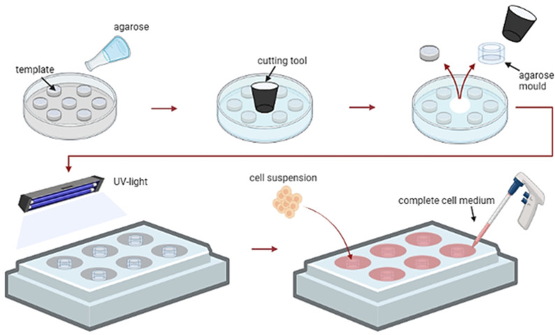
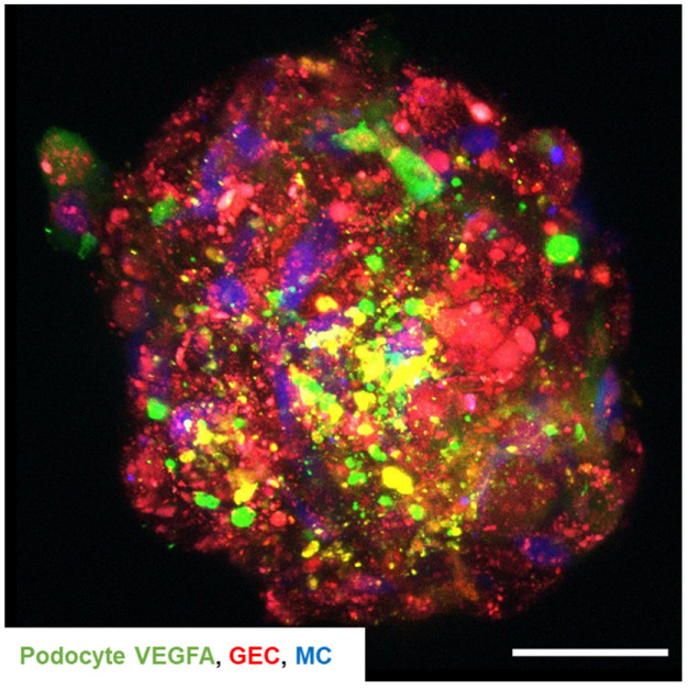
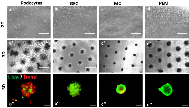
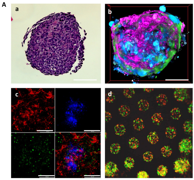
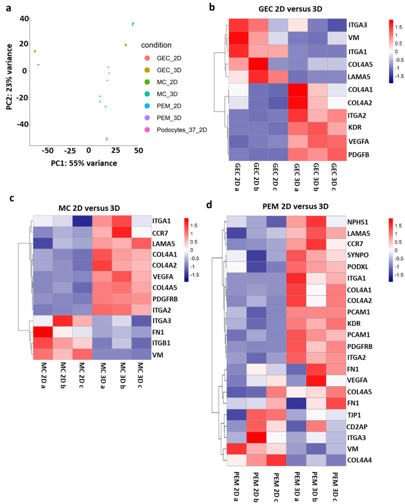
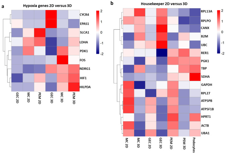
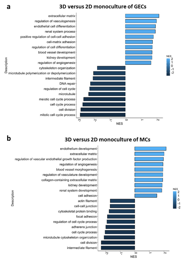
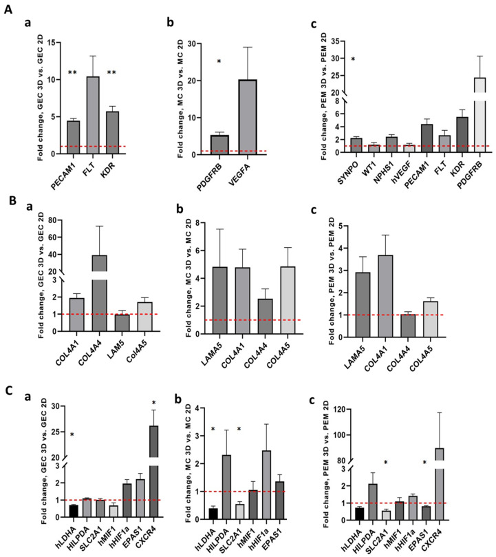
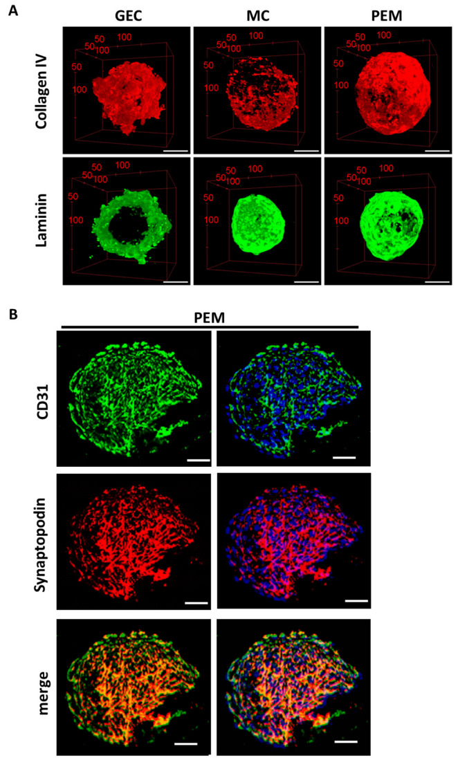
Signaling-pathway analyses and the investigation of gene responses to different stimuli are usually performed in 2D monocultures. However, within the glomerulus, cells grow in 3D and are involved in direct and paracrine interactions with different glomerular cell types. Thus, the results from 2D monoculture experiments must be taken with caution. We cultured glomerular endothelial cells, podocytes and mesangial cells in 2D/3D monocultures and 2D/3D co-cultures and analyzed cell survival, self-assembly, gene expression, cell-cell interaction, and gene pathways using live/dead assay, time-lapse analysis, bulk-RNA sequencing, qPCR, and immunofluorescence staining. Without any need for scaffolds, 3D glomerular co-cultures self-organized into spheroids. Podocyte- and glomerular endothelial cell-specific markers and the extracellular matrix were increased in 3D co-cultures compared to 2D co-cultures. Housekeeping genes must be chosen wisely, as many genes used for the normalization of gene expression were themselves affected in 3D culture conditions. The transport of podocyte-derived VEGFA to glomerular endothelial cells confirmed intercellular crosstalk in the 3D co-culture models. The enhanced expression of genes important for glomerular function in 3D, compared to 2D, questions the reliability of currently used 2D monocultures. Hence, glomerular 3D co-cultures might be more suitable in the study of intercellular communication, disease modelling and drug screening ex vivo.
信号通路分析以及对不同刺激下基因反应的研究通常在 2D 单层培养中进行。然而,在肾小球中,细胞在 3D 中生长,并与不同的肾小球细胞类型进行直接和旁分泌相互作用。因此,必须谨慎对待来自 2D 单层培养实验的结果。我们在 2D/3D 单层和 2D/3D 共培养中培养肾小球内皮细胞、足细胞和系膜细胞,并使用活/死检测、延时分析、批量 RNA 测序、qPCR 和免疫荧光染色分析细胞存活、自我组装、基因表达、细胞-细胞相互作用和基因通路。在不需要支架的情况下,3D 肾小球共培养物自发组织成球体。与 2D 共培养物相比,3D 共培养物中足细胞和肾小球内皮细胞特异性标志物和细胞外基质增加。管家基因必须谨慎选择,因为许多用于基因表达归一化的基因本身在 3D 培养条件下受到影响。足细胞衍生的 VEGFA 向肾小球内皮细胞的转运证实了 3D 共培养模型中的细胞间串扰。与 2D 相比,3D 中与肾小球功能重要的基因的表达增强,对目前使用的 2D 单层培养的可靠性提出了质疑。因此,肾小球 3D 共培养可能更适合于细胞间通讯、疾病建模和药物筛选的体外研究。