Department of Pathology, Faculty of Medicine, Aristotle University of Thessaloniki, Thessaloniki, Greece.
European Society of Pathology, Brussels, Belgium.
Virchows Arch. 2023 Dec;483(6):775-786. doi: 10.1007/s00428-023-03581-y. Epub 2023 Jul 4.
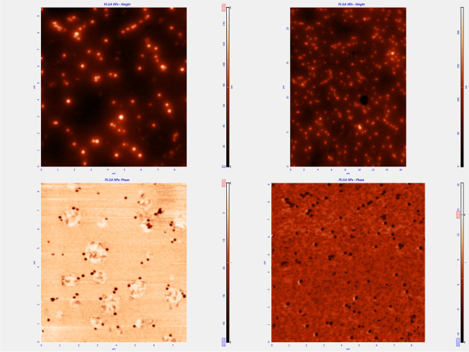
Over the last years, there has been an increasing number of proposals for the use of nanomaterials in medicine. The safety of novel technologies must be verified, prior to their clinical application. Pathology has much to contribute towards this end. In this study, we compared the in vivo toxicity effects of poly- (lactic-co-glycolic acid) nanoparticles with and without chitosan shell. Both nanoparticle types were loaded with curcumin. The nanoparticles were assessed in vitro for potential cytotoxicity with cell viability studies. For the in vivo test, 36 adult Wistar rats were used, four of which were the control group. The remaining 32 were divided into 2 groups, each of which was administered differentially coated drug carriers: (A) nanoparticles without chitosan coating and (B) nanoparticles with chitosan coating. For both groups, the subcutaneous route was used for administration. Each group was further divided into 2 sub-groups of 8 animals each. The animals of the first sub-groups were sacrificed 24 h after the injection and those of the second on the 7th day. The control group was also divided into 2 subgroups of 2 animals each. At the appointed post-administrative date, the rats were sacrificed, and specimens from the brain, liver, kidneys, heart, stomach, lungs, and from the skin at the injection site were collected and studied histopathologically. The evaluation of both in vitro and in vivo testing shows that nanoparticles with chitosan have significantly less, if any, toxic effects compared to those without chitosan.
近年来,越来越多的人提议将纳米材料应用于医学领域。在将新技术应用于临床之前,必须对其安全性进行验证。病理学为此做出了很大的贡献。在这项研究中,我们比较了具有壳聚糖壳和不具有壳聚糖壳的聚(乳酸-共-乙醇酸)纳米粒子的体内毒性效应。这两种纳米粒子类型都负载了姜黄素。通过细胞活力研究评估了纳米粒子的体外潜在细胞毒性。在体内试验中,使用了 36 只成年 Wistar 大鼠,其中 4 只为对照组。其余 32 只大鼠分为 2 组,每组分别给予不同包被的药物载体:(A)无壳聚糖包被的纳米粒子和(B)有壳聚糖包被的纳米粒子。对于这两组,均采用皮下途径给药。每组进一步分为 2 个亚组,每组 8 只动物。第一亚组动物在注射后 24 小时处死,第二亚组动物在第 7 天处死。对照组也分为 2 个亚组,每组 2 只动物。在指定的给药后日期,处死大鼠,采集脑、肝、肾、心、胃、肺和注射部位皮肤的标本,并进行组织病理学研究。体外和体内试验的评估表明,具有壳聚糖的纳米粒子的毒性作用明显小于不具有壳聚糖的纳米粒子。