Department of Neurology, State Key Laboratory of Complex Severe and Rare Disease, Peking Union Medical College Hospital, Chinese Academy of Medical Science and Peking Union Medical College, Beijing 100730, China.
Department of Radiology, Peking Union Medical College Hospital, Chinese Academy of Medical Sciences and Peking Union Medical College, Beijing 100730, China.
Chin Med J (Engl). 2024 Apr 5;137(7):830-836. doi: 10.1097/CM9.0000000000002785. Epub 2023 Jul 7.
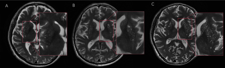
In the clinic, practitioners encounter many patients with an abnormal pattern of dense punctate magnetic resonance imaging (MRI) signal in the basal ganglia, a phenomenon known as "cheese sign". This sign is reported as common in cerebrovascular diseases, dementia, and old age. Recently, cheese sign has been speculated to consist of dense perivascular space (PVS). This study aimed to assess the lesion types of cheese sign and analyze the correlation between this sign and vascular disease risk factors.
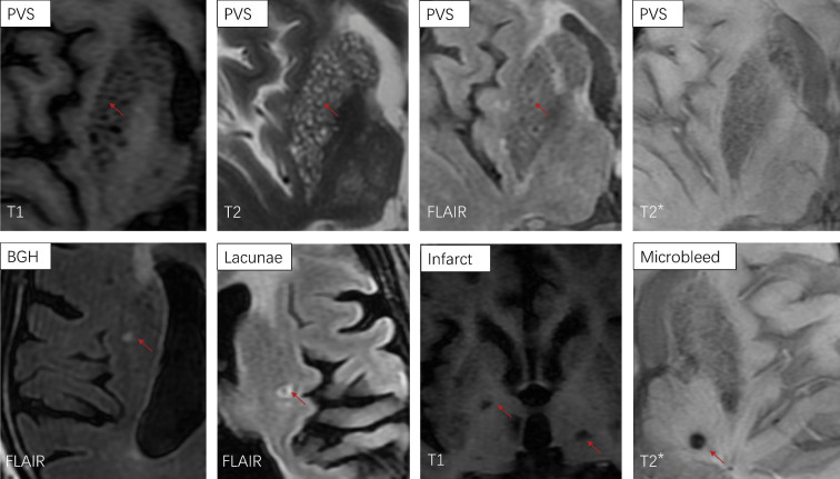
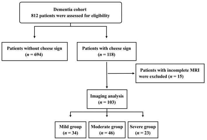
A total of 812 patients from Peking Union Medical College Hospital (PUMCH) dementia cohort were enrolled. We analyzed the relationship between cheese sign and vascular risk. For assessing cheese sign and defining its degree, the abnormal punctate signals were classified into basal ganglia hyperintensity (BGH), PVS, lacunae/infarctions and microbleeds, and counted separately. Each type of lesion was rated on a four-level scale, and then the sum was calculated; this total was defined as the cheese sign score. Fazekas and Age-Related White Matter Changes (ARWMC) scores were used to evaluate the paraventricular, deep, and subcortical gray/white matter hyperintensities.
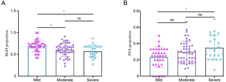
A total of 118 patients (14.5%) in this dementia cohort were found to have cheese sign. Age (odds ratio [OR]: 1.090, 95% confidence interval [CI]: 1.064-1.120, P <0.001), hypertension (OR: 1.828, 95% CI: 1.123-2.983, P = 0.014), and stroke (OR: 1.901, 95% CI: 1.092-3.259, P = 0.025) were risk factors for cheese sign. There was no significant relationship between diabetes, hyperlipidemia, and cheese sign. The main components of cheese sign were BGH, PVS, and lacunae/infarction. The proportion of PVS increased with cheese sign severity.
The risk factors for cheese sign were hypertension, age, and stroke. Cheese sign consists of BGH, PVS, and lacunae/infarction.
在临床中,医生会遇到许多基底节区存在异常点状致密磁共振成像(MRI)信号的患者,这种现象被称为“奶酪征”。该征象被报道在脑血管病、痴呆和老年人群中较为常见。最近,有研究推测“奶酪征”由致密的血管周围间隙(PVS)组成。本研究旨在评估“奶酪征”的病变类型,并分析该征象与血管疾病危险因素之间的关系。
本研究纳入了北京协和医院痴呆队列的 812 例患者。我们分析了“奶酪征”与血管危险因素之间的关系。为了评估“奶酪征”并对其严重程度进行分析,我们将异常点状信号分为基底节高信号(BGH)、PVS、腔隙/梗死和微出血,并分别进行计数。每种类型的病变均进行四级评分,然后进行总和,定义为“奶酪征”评分。Fazekas 评分和年龄相关性脑白质改变(ARWMC)评分用于评估脑室周围、深部和皮质下灰质/白质高信号。
在该痴呆队列中,共有 118 例(14.5%)患者存在“奶酪征”。年龄(比值比 [OR]:1.090,95%置信区间 [CI]:1.064-1.120,P <0.001)、高血压(OR:1.828,95% CI:1.123-2.983,P = 0.014)和卒中(OR:1.901,95% CI:1.092-3.259,P = 0.025)是“奶酪征”的危险因素。糖尿病和高脂血症与“奶酪征”无显著相关性。“奶酪征”的主要成分是 BGH、PVS 和腔隙/梗死。“奶酪征”严重程度与 PVS 比例呈正相关。
“奶酪征”的危险因素包括高血压、年龄和卒中。“奶酪征”由 BGH、PVS 和腔隙/梗死组成。