Department of Cancer Biology and Molecular Medicine, Beckman Research Institute, City of Hope Comprehensive Cancer Center, Duarte, California, USA.
Cardiology Division and Corrigan Minehan Heart Center, Massachusetts General Hospital, Harvard Medical School, Boston, Massachusetts, USA.
J Extracell Vesicles. 2023 Jul;12(7):e12346. doi: 10.1002/jev2.12346.
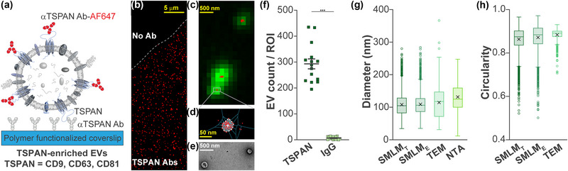
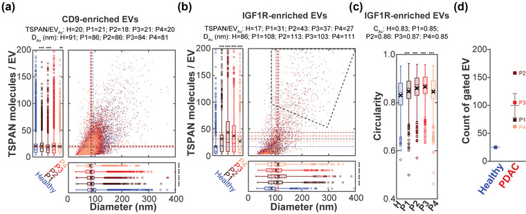
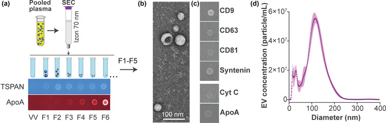
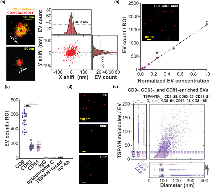
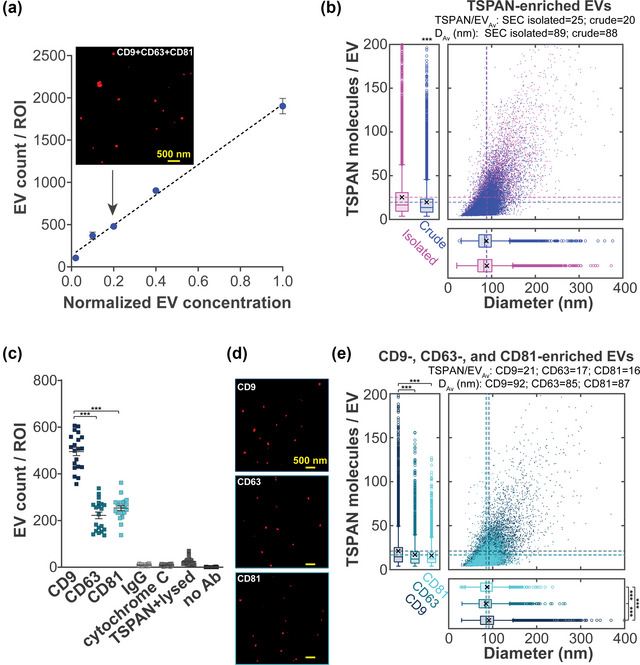
Extracellular vesicles (EVs) and their cargo constitute novel biomarkers. EV subpopulations have been defined not only by abundant tetraspanins (e.g., CD9, CD63 and CD81) but also by specific markers derived from their source cells. However, it remains a challenge to robustly isolate and characterize EV subpopulations. Here, we combined affinity isolation with super-resolution imaging to comprehensively assess EV subpopulations from human plasma. Our Single Extracellular VEsicle Nanoscopy (SEVEN) assay successfully quantified the number of affinity-isolated EVs, their size, shape, molecular tetraspanin content, and heterogeneity. The number of detected tetraspanin-enriched EVs positively correlated with sample dilution in a 64-fold range (for SEC-enriched plasma) and a 50-fold range (for crude plasma). Importantly, SEVEN robustly detected EVs from as little as ∼0.1 μL of crude plasma. We further characterized the size, shape and molecular tetraspanin content (with corresponding heterogeneities) for CD9-, CD63- and CD81-enriched EV subpopulations. Finally, we assessed EVs from the plasma of four pancreatic ductal adenocarcinoma patients with resectable disease. Compared to healthy plasma, CD9-enriched EVs from patients were smaller while IGF1R-enriched EVs from patients were larger, rounder and contained more tetraspanin molecules, suggestive of a unique pancreatic cancer-enriched EV subpopulation. This study provides the method validation and demonstrates that SEVEN could be advanced into a platform for characterizing both disease-associated and organ-associated EV subpopulations.
细胞外囊泡 (EVs) 及其 cargo 构成了新型生物标志物。EV 亚群不仅通过丰富的四跨膜蛋白(例如 CD9、CD63 和 CD81)定义,还可以通过其来源细胞的特异性标志物定义。然而,稳健地分离和表征 EV 亚群仍然是一个挑战。在这里,我们将亲和分离与超分辨率成像相结合,全面评估来自人血浆的 EV 亚群。我们的单个细胞外囊泡纳米显微镜 (SEVEN) 检测成功地定量了亲和分离的 EV 数量、它们的大小、形状、分子四跨膜蛋白含量和异质性。检测到的富含四跨膜蛋白的 EV 数量与样品稀释度呈正相关,在 64 倍范围内(对于 SEC 富集的血浆)和 50 倍范围内(对于粗血浆)。重要的是,SEVEN 能够从仅约 0.1 μL 的粗血浆中稳健地检测到 EV。我们进一步表征了 CD9、CD63 和 CD81 富集 EV 亚群的大小、形状和分子四跨膜蛋白含量(及其相应的异质性)。最后,我们评估了四位可切除疾病的胰腺导管腺癌患者的血浆中的 EV。与健康血浆相比,来自患者的 CD9 富集 EV 较小,而来自患者的 IGF1R 富集 EV 较大、较圆,并且含有更多的四跨膜蛋白分子,提示存在独特的胰腺癌症富集 EV 亚群。这项研究提供了方法验证,并表明 SEVEN 可以推进到用于表征疾病相关和器官相关 EV 亚群的平台。