Department of Radiology, The First Affiliated Hospital of Xi'an Jiaotong University, Xi'an, China.
Shaanxi Provincial Key Laboratory of Clinic Genetics, Fourth Military Medical University, Xi'an, China.
BMC Med. 2023 Jul 10;21(1):250. doi: 10.1186/s12916-023-02963-y.
Inflammation has been implicated in the pathology of schizophrenia and may cause neuronal cell death and dendrite loss. Neuroimaging studies have highlighted longitudinal brain structural changes in patients with schizophrenia, yet it is unclear whether this is related to inflammation. We aim to address this question, by relating brain structural changes with the transcriptional profile of inflammation markers in the early stage of schizophrenia.
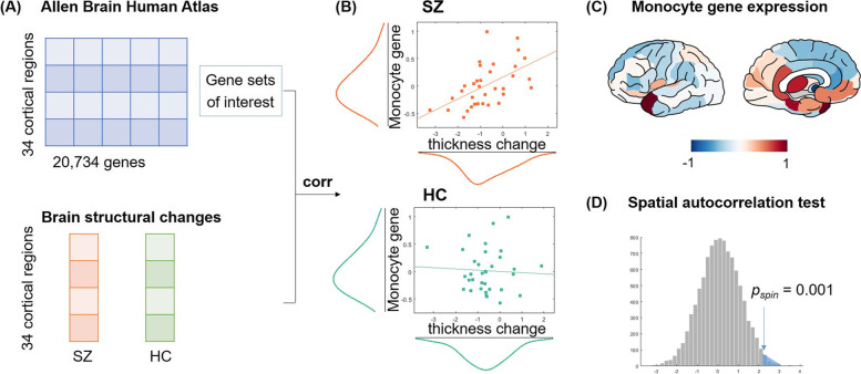
Thirty-eight patients with first-episode schizophrenia and 51 healthy controls were included. High-resolution T1-weighted magnetic resonance imaging (MRI) and clinical assessments were performed at baseline and 2 ~ 6 months follow-up for all subjects. Changes in the brain structure were analyzed using surface-based morphological analysis and correlated with the expression of immune cells-related gene sets of interest reported by previous reviews. Transcriptional data were retrieved from the Allen Human Brain Atlas. Furthermore, we examined the brain structural changes and peripheral inflammation markers in association with behavioral symptoms and cognitive functioning in patients.
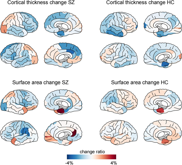
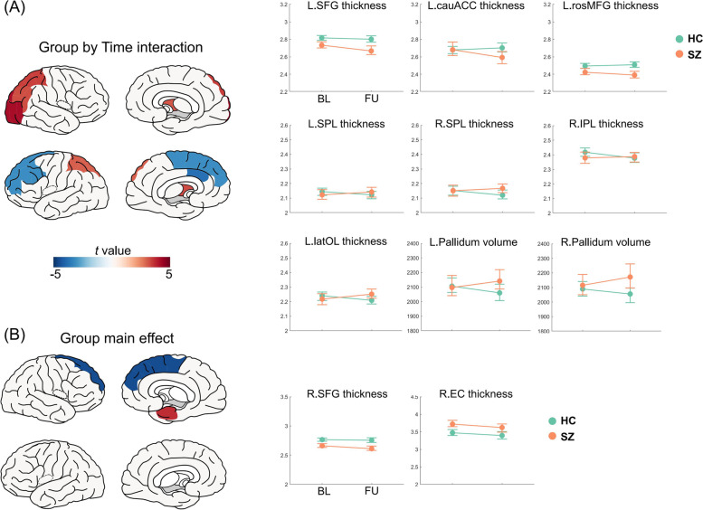
Patients exhibited accelerated cortical thickness decrease in the left frontal cortices, less decrease or an increase in the superior parietal lobule and right lateral occipital lobe, and increased volume in the bilateral pallidum, compared with controls. Changes in cortical thickness correlated with the transcriptional level of monocyte across cortical regions in patients (r = 0.54, p < 0.01), but not in controls (r = - 0.05, p = 0.76). In addition, cortical thickness change in the left superior parietal lobule positively correlated with changes in digital span-backward test scores in patients.
Patients with schizophrenia exhibit regional-specific cortical thickness changes in the prefrontal and parietooccipital cortices, which is related to their cognitive impairment. Inflammation may be an important factor contributing to cortical thinning in first-episode schizophrenia. Our findings suggest that the immunity-brain-behavior association may play a crucial role in the pathogenesis of schizophrenia.
炎症与精神分裂症的病理学有关,可能导致神经元细胞死亡和树突丢失。神经影像学研究强调了精神分裂症患者的纵向脑结构变化,但尚不清楚这是否与炎症有关。我们旨在通过将炎症标志物的转录谱与精神分裂症早期的脑结构变化相关联来解决这个问题。
纳入 38 例首发精神分裂症患者和 51 例健康对照者。所有受试者均在基线和 2 至 6 个月时进行高分辨率 T1 加权磁共振成像(MRI)和临床评估。使用基于表面的形态分析来分析脑结构变化,并与以前综述报道的与免疫细胞相关的基因集的表达相关联。转录数据从艾伦人类大脑图谱中检索。此外,我们在患者中检查了脑结构变化与外周炎症标志物与行为症状和认知功能的关联。
与对照组相比,患者的左侧额皮质表现出皮质厚度的加速下降,顶叶上回和右侧外侧枕叶的皮质厚度减少或增加,双侧苍白球体积增加。与对照组(r = -0.05,p = 0.76)相比,患者的皮质厚度变化与皮质区域的单核细胞转录水平相关(r = 0.54,p < 0.01)。此外,左侧顶叶上回的皮质厚度变化与患者数字广度回溯测试评分的变化呈正相关。
首发精神分裂症患者表现出前额叶和顶枕叶的皮质厚度的区域特异性变化,这与他们的认知障碍有关。炎症可能是首发精神分裂症皮质变薄的一个重要因素。我们的研究结果表明,免疫-脑-行为的关联可能在精神分裂症的发病机制中起着关键作用。