Department of Emergency and General Internal Medicine, Fujita Health University School of Medicine, 1-98 Dengakugakubo, Kutsukakecho, Toyoake, Aichi, 470-1192, Japan.
Division of Intensive Care Medicine, Department of Internal Medicine, Okinawa Prefectural Chubu Hospital, Uruma, Japan.
Crit Care. 2023 Jul 10;27(1):278. doi: 10.1186/s13054-023-04557-9.
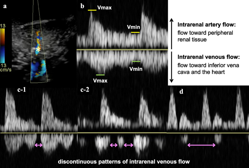
Intrarenal venous flow (IRVF) patterns assessed using Doppler renal ultrasonography are real-time bedside visualizations of renal vein hemodynamics. Although this technique has the potential to detect renal congestion during sepsis resuscitation, there have been few studies on this method. We aimed to examine the relationship between IRVF patterns, clinical parameters, and outcomes in critically ill adult patients with sepsis. We hypothesized that discontinuous IRVF was associated with elevated central venous pressure (CVP) and subsequent acute kidney injury (AKI) or death.
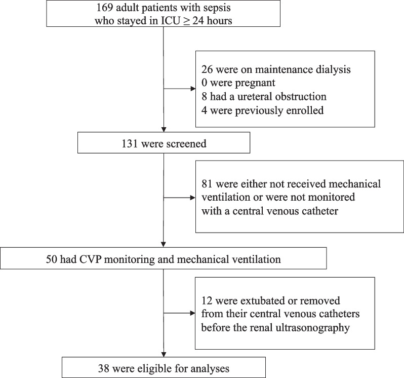
We conducted a prospective observational study in two tertiary-care hospitals, enrolling adult patients with sepsis who stayed in the intensive care unit for at least 24 h, had central venous catheters placed, and received invasive mechanical ventilation. Renal ultrasonography was performed at a single time point at the bedside after sepsis resuscitation, and IRVF patterns (discontinuous vs. continuous) were confirmed by a blinded assessor. The primary outcome was CVP obtained at the time of renal ultrasonography. We also repeatedly assessed a composite of Kidney Disease Improving Global Outcomes of Stage 3 AKI or death over the course of a week as a secondary outcome. The association of IRVF patterns with CVP was examined using Student's t-test (primary analysis) and that with composite outcomes was assessed using a generalized estimating equation analysis, to account for intra-individual correlations. A sample size of 32 was set in order to detect a 5-mmHg difference in CVP between IRVF patterns.
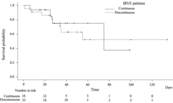
Of the 38 patients who met the eligibility criteria, 22 (57.9%) showed discontinuous IRVF patterns that suggested blunted renal venous flow. IRVF patterns were not associated with CVP (discontinuous flow group: mean 9.24 cm HO [standard deviation: 3.19], continuous flow group: 10.65 cm HO [standard deviation: 2.53], p = 0.154). By contrast, the composite outcome incidence was significantly higher in the discontinuous IRVF pattern group (odds ratio: 9.67; 95% confidence interval: 2.13-44.03, p = 0.003).
IRVF patterns were not associated with CVP but were associated with subsequent AKI in critically ill adult patients with sepsis. IRVF may be useful for capturing renal congestion at the bedside that is related to clinical patient outcomes.
使用多普勒肾超声评估的肾内静脉血流 (IRVF) 模式是肾静脉血液动力学的实时床边可视化。尽管该技术有可能在脓毒症复苏期间检测到肾淤血,但对此方法的研究甚少。我们旨在检查危重病成人脓毒症患者的 IRVF 模式、临床参数和结局之间的关系。我们假设不连续的 IRVF 与中心静脉压 (CVP) 升高以及随后的急性肾损伤 (AKI) 或死亡有关。
我们在两家三级保健医院进行了一项前瞻性观察性研究,纳入了至少在重症监护病房停留 24 小时、放置中心静脉导管和接受有创机械通气的脓毒症成年患者。在脓毒症复苏后在床边进行单次肾脏超声检查,并由盲法评估员确认 IRVF 模式(不连续与连续)。主要结局是在进行肾脏超声检查时获得的 CVP。我们还在一周的时间内反复评估了肾脏病改善全球结局 3 期 AKI 或死亡的综合指标作为次要结局。使用学生 t 检验(主要分析)检查 IRVF 模式与 CVP 的关系,并使用广义估计方程分析评估与综合结局的关系,以考虑个体内相关性。为了检测 IRVF 模式之间的 CVP 差异 5mmHg,设定了 32 的样本量。
在符合入选标准的 38 名患者中,22 名(57.9%)表现出不连续的 IRVF 模式,提示肾静脉血流减弱。IRVF 模式与 CVP 无关(不连续血流组:平均 9.24cmH2O[标准差:3.19],连续血流组:10.65cmH2O[标准差:2.53],p=0.154)。相比之下,不连续 IRVF 模式组的复合结局发生率显著更高(比值比:9.67;95%置信区间:2.13-44.03,p=0.003)。
IRVF 模式与 CVP 无关,但与危重病成人脓毒症患者的后续 AKI 有关。IRVF 可用于床边捕捉与临床患者结局相关的肾淤血。