Zlatkov Boyan, Vergilov Vladislav, Pérez Santa-Rita José Vicente, Baixeras Joaquín
Institute of Biodiversity and Ecosystem Research, Bulgarian Academy of Sciences, 1 Tsar Osvoboditel Blvd., 1000, Sofia, Bulgaria.
National Museum of Natural History, Bulgarian Academy of Sciences, 1 Tsar Osvoboditel Blvd., 1000, Sofia, Bulgaria.
Front Zool. 2023 Jul 11;20(1):22. doi: 10.1186/s12983-023-00500-4.
The process of copulation in Lepidoptera is understudied and poorly understood from a functional perspective. The purpose of the present paper is to study the interaction of the male and female genitalia of Tortrix viridana Linnaeus, 1758 via three-dimensional models of pairs fixed during copulation. Other techniques (confocal laser scanning microscopy, scanning electron microscopy and histology) were used to clarify the role of the organs involved in the process.
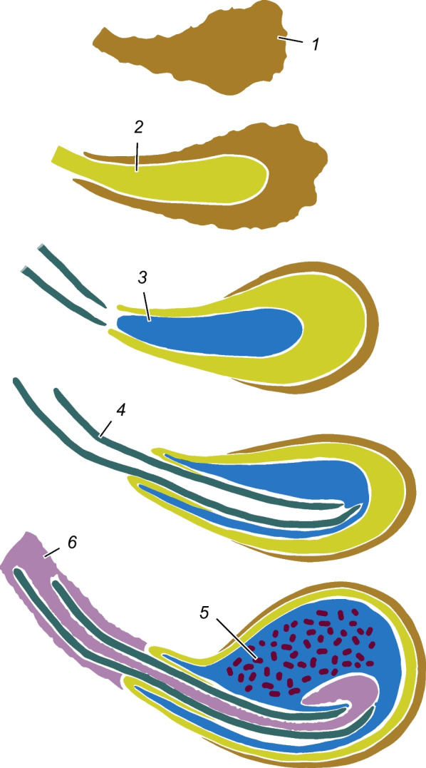
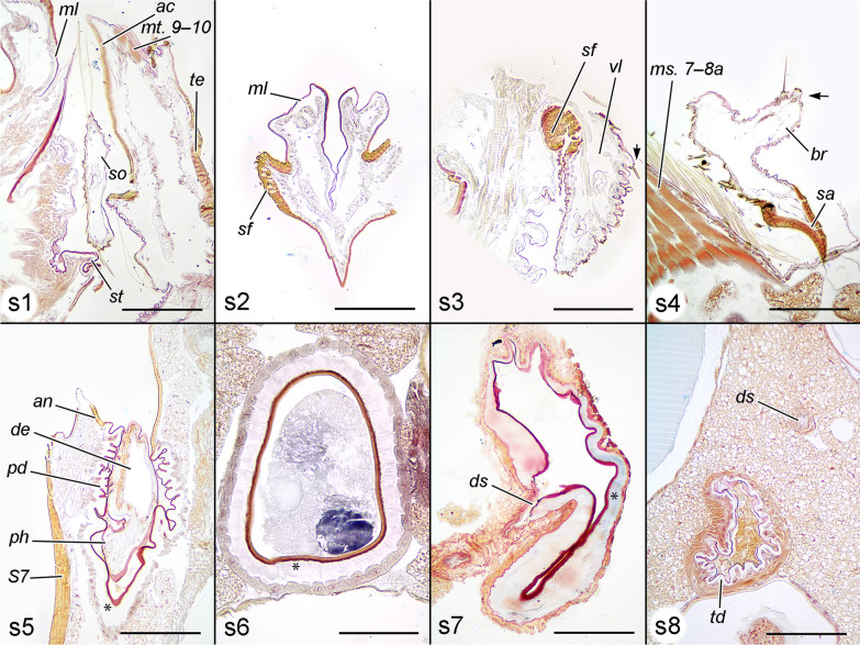
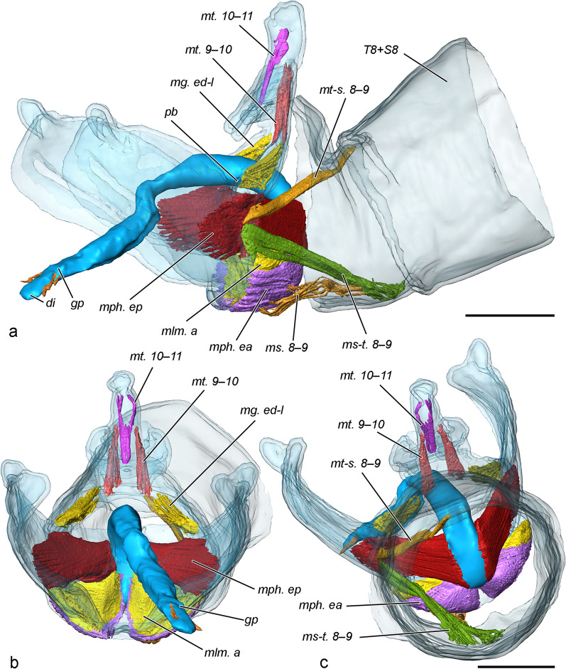
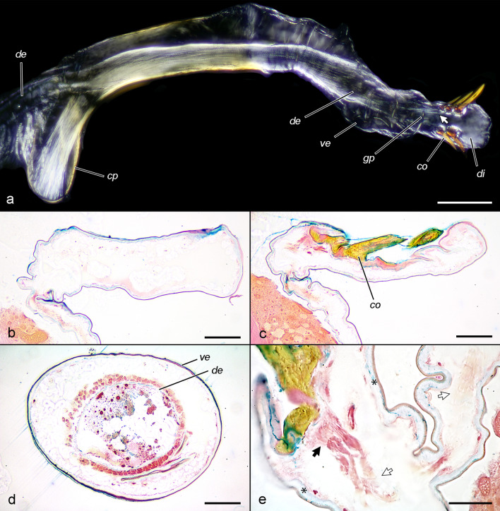
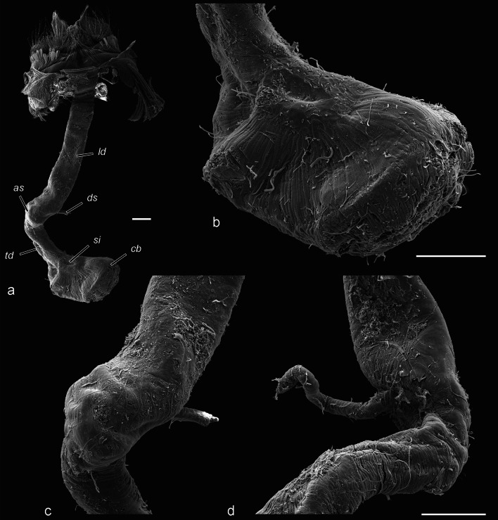
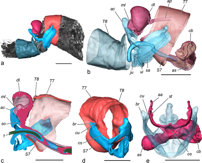
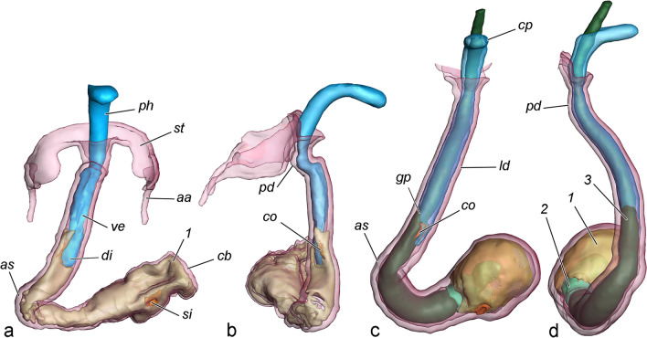
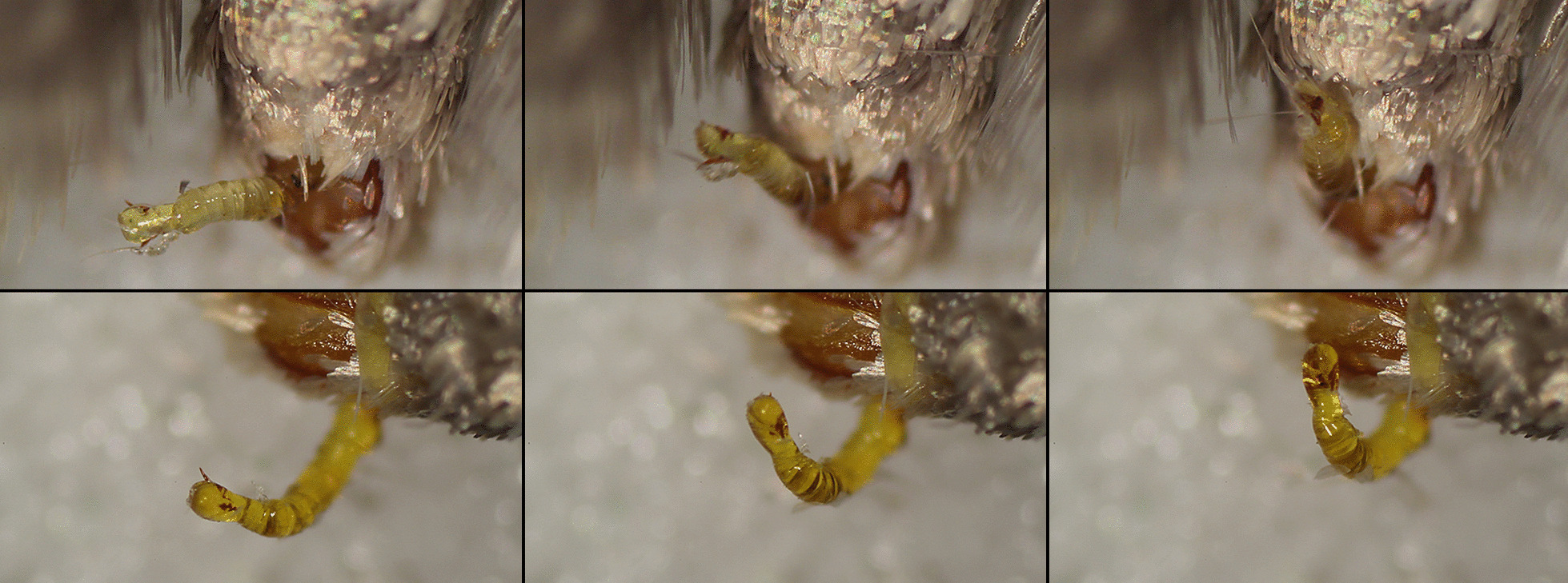
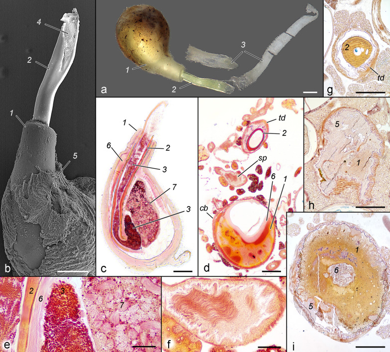
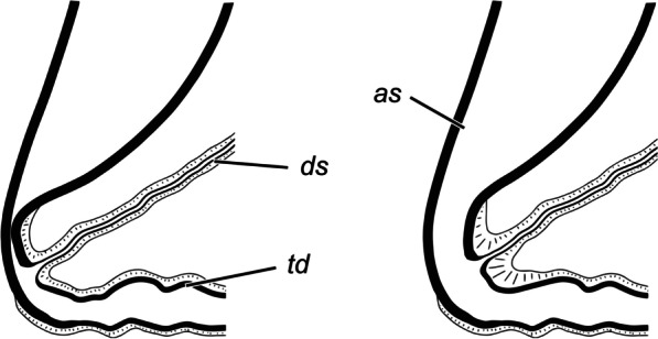
Three-dimensional models based on micro-CT scanned copulating pairs were generated allowing visualisation of the position of the male and female counterparts, spatial changes during copulation, and the skeleto-muscular apparatus involved in the process. The male genitalia and their musculature are simplified in comparison with other lineages of the family, but the opposite is true for the female genitalia. The attachment of the couple is achieved only through flexion of the valvae, clasping the large and sclerotised sternite 7 of the female. The anal cone and socii of the male are in contact with certain parts of the anal papillae and the sterigma of the female. The long tubular vesica is inserted into the narrow posterior part of the ductus bursae. Its eversion is achieved by an increase in haemolymph pressure. A possible mechanism of stimulation of the female via pulsations of the diverticulum of the vesica was discovered. A compressed sclerotised area of the ductus bursae putatively serves as a valve controlling the transfer of ejaculated materials. Copulation progresses through two phases: in the first the vesica and its diverticulum are inflated by haemolymph, and in the second the diverticulum is not inflated, and the vesica is occupied by viscous ejaculated material. The formation of the multilayered spermatophore was observed, and we discovered that sperm is transferred very late in the copulation process.
Copulation process in Lepidoptera is studied for the first time with three-dimensional reconstructions of couples of Tortrix viridana, used as a model species. The internal genitalia is the scenario of multiple interactions between male and female, but the external remain static. A possible mechanism of stimulation of the female internal copulation organs is proposed.
鳞翅目昆虫的交配过程研究较少,从功能角度理解也很有限。本文旨在通过对交配时固定的雌雄配对的三维模型,研究1758年林奈命名的绿卷蛾(Tortrix viridana)雌雄生殖器的相互作用。还运用了其他技术(共聚焦激光扫描显微镜、扫描电子显微镜和组织学)来阐明参与该过程的器官的作用。
基于微计算机断层扫描(micro-CT)扫描的交配配对生成了三维模型,从而能够可视化雌雄对应器官的位置、交配过程中的空间变化以及参与该过程的骨骼肌肉结构。与该科的其他谱系相比,雄性生殖器及其肌肉组织较为简化,但雌性生殖器则相反。雌雄的连接仅通过瓣的弯曲来实现,瓣扣住雌性较大且硬化的第7腹节腹板。雄性的肛锥和抱器与雌性肛乳头和气门的某些部分接触。长管状的贮精囊插入囊导管的狭窄后部。贮精囊的外翻通过血淋巴压力的增加来实现。发现了一种通过贮精囊憩室的脉动刺激雌性的可能机制。囊导管的一个压缩硬化区域可能起到控制射精物质转移的瓣膜作用。交配过程分为两个阶段:第一阶段,贮精囊及其憩室被血淋巴充盈;第二阶段,憩室未充盈,贮精囊被粘性射精物质占据。观察到多层精包的形成,并且我们发现精子在交配过程的后期才转移。
首次以绿卷蛾作为模式物种,通过对其雌雄配对的三维重建来研究鳞翅目的交配过程。内部生殖器是雌雄之间多种相互作用的场所,但外部保持静止。提出了一种刺激雌性内部交配器官的可能机制。