• 文献检索
  • 文档翻译
  • 深度研究
  • 学术资讯
  • Suppr Zotero 插件Zotero 插件
  • 邀请有礼
  • 套餐&价格
  • 历史记录
应用&插件
Suppr Zotero 插件Zotero 插件浏览器插件Mac 客户端Windows 客户端微信小程序
定价
高级版会员购买积分包购买API积分包
服务
文献检索文档翻译深度研究API 文档MCP 服务
关于我们
关于 Suppr公司介绍联系我们用户协议隐私条款
关注我们

Suppr 超能文献

核心技术专利:CN118964589B侵权必究
粤ICP备2023148730 号-1Suppr @ 2026

文献检索

告别复杂PubMed语法,用中文像聊天一样搜索,搜遍4000万医学文献。AI智能推荐,让科研检索更轻松。

立即免费搜索

文件翻译

保留排版,准确专业,支持PDF/Word/PPT等文件格式,支持 12+语言互译。

免费翻译文档

深度研究

AI帮你快速写综述,25分钟生成高质量综述,智能提取关键信息,辅助科研写作。

立即免费体验

定量计算机断层扫描中C1-C2骨密度的区域差异及其临床意义。

Regional variations in C1-C2 bone density on quantitated computed tomography and clinical implications.

作者信息

Baisden Jamie L, Varghese Vicky, Banerjee Anjishnu, Yoganandan Narayan

机构信息

Department of Neurosurgery, Medical College of Wisconsin, 8701 Watertown Plank Rd, Milwaukee, WI 53226, United States.

Department of Orthopedics, Skokie Hospital, NorthShore University Health System, 9600 Gross Point Rd, Skokie, IL 60076, United States.

出版信息

N Am Spine Soc J. 2023 May 11;14:100228. doi: 10.1016/j.xnsj.2023.100228. eCollection 2023 Jun.

DOI:10.1016/j.xnsj.2023.100228
PMID:37440985
原文链接:https://pmc.ncbi.nlm.nih.gov/articles/PMC10333715/
Abstract

BACKGROUND

Our elderly population is growing and the number of spine fractures in the elderly is also growing. The elderly population in general may be considered as poor surgical candidates experience a high rate of fractures at C1 and C2 compared with the general population. Nonoperative management of upper cervical fractures is not benign as there is a high nonunion rate for both C1 and C2 fractures in the elderly, and orthosis compliance is often suboptimal, or complicated by skin breakdown. The optimal technique for upper cervical stabilization in the elderly may be different than in younger populations as the bone quality is inferior in the elderly. The objective of this basic science study is to determine whether the bone mineral density (BMD) of C1 and C2 vary by region, and if this is a gender difference in this elderly age group.

METHODS

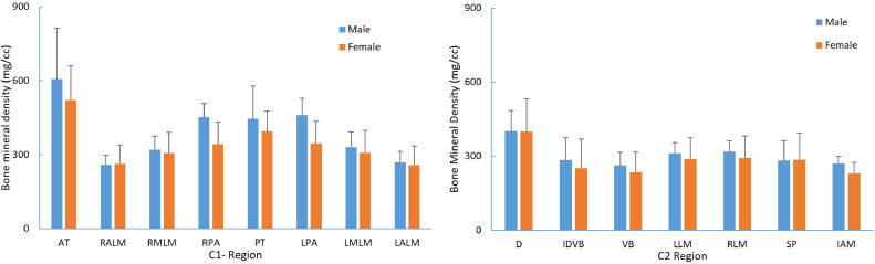
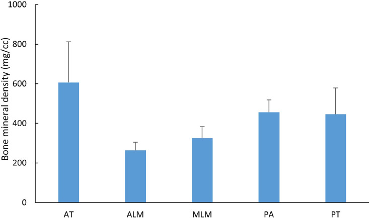
Twenty cadaveric spines from 45 to 83 years of age were used to obtain BMD using quantitated computed tomography (QCT). BMD was measured using a QCT. For C1, 8 regions were determined: anterior tubercle, bilateral anterior and medial lateral masses, bilateral posterior arches, and posterior tubercle. For C2, 7 regional BMDs were determined: top of odontoid, base of odontoid-body interface, mid body, bilateral lateral masses, anterior inferior body near the discs space, and the C2 spinous process.

RESULTS

The BMD was greatest at the C1 anterior tubercle (564.4±175.8 mg/cm) and C1 posterior ring (420.8±110.2 mg/cm), and least at the anterior and medial lateral masses (262.8±59.5 mg/cm, 316.9±72.6 mg/cm). At C2 QCT BMD was greatest at the top of the dens (400.6±107.9 mg/cm) decreasing down through the odontoid-C2 body junction (267.8±103.5 mg/cm) and least in the mid C2 body 249.1±68.8 mg/cm). The posterior arch of C1 and the spinous process of C2 had higher BMD's 420.8±110.2 mg/cm and 284.1±93.0 mg/cm, respectively. A high correlation was observed between the BMD at the interface of the dens-vertebral body with the vertebral body with a Pearson correlation coefficient of 0.86. The BMD of the top of dens was significantly higher (p<.05) than all the regions in C2.

CONCLUSIONS

Regional and segmental BMD variations at C1 and C2 have clinical implications for surgical constructs in the elderly population. Given the higher BMDs of the C1 and C2 spinous process and posterior arches, consideration should be given to incorporate these areas using various C1-C2 wiring techniques. In the elderly, lateral masses particularly at C1 with lower BMD may result in potential screw loosening and nonunion in this age group. Old-school wiring techniques have a track record of efficacy and safety with less blood loss, reduced operative time, reduced X-ray exposure, and should be considered in the elderly as a primary stabilization technique or a belt-over suspenders approach based on regional variations in BMD in the elderly.

摘要

背景

我们的老年人口正在增长,老年人脊柱骨折的数量也在增加。一般来说,老年人群体可能被视为手术候选不佳者,与普通人群相比,他们在C1和C2处的骨折发生率较高。老年患者上颈椎骨折的非手术治疗并非无害,因为老年患者C1和C2骨折的不愈合率很高,而且矫形器的依从性往往不理想,或者会并发皮肤破损。由于老年人的骨质较差,老年人上颈椎稳定的最佳技术可能与年轻人群体不同。这项基础科学研究的目的是确定C1和C2的骨密度(BMD)是否因区域而异,以及在这个老年年龄组中是否存在性别差异。

方法

使用20具年龄在45至83岁之间的尸体脊柱,通过定量计算机断层扫描(QCT)获得骨密度。使用QCT测量骨密度。对于C1,确定了8个区域:前结节、双侧前外侧块和后外侧块、双侧后弓以及后结节。对于C2,确定了7个区域的骨密度:齿突顶部、齿突-椎体界面底部、椎体中部、双侧侧块、椎间盘间隙附近的椎体前下部以及C2棘突。

结果

C1前结节(564.4±175.8mg/cm)和C1后环(420.8±110.2mg/cm)的骨密度最高,前外侧块和后外侧块(262.8±59.5mg/cm,316.9±72.6mg/cm)的骨密度最低。在C2,QCT骨密度在齿突顶部(400.6±107.9mg/cm)最高,向下穿过齿突-C2椎体交界处(267.8±103.5mg/cm)逐渐降低,在C2椎体中部最低(249.1±68.8mg/cm)。C1后弓和C2棘突的骨密度较高,分别为420.8±110.2mg/cm和284.1±93.0mg/cm。在齿突-椎体界面处的骨密度与椎体骨密度之间观察到高度相关性,皮尔逊相关系数为0.86。齿突顶部的骨密度显著高于C2的所有区域(p<0.05)。

结论

C1和C2的区域和节段性骨密度变化对老年人群体的手术构建具有临床意义。鉴于C1和C2棘突及后弓的骨密度较高,应考虑使用各种C1-C2钢丝技术将这些区域纳入。在老年人中,特别是C1处骨密度较低的侧块可能导致该年龄组潜在的螺钉松动和不愈合。传统的钢丝技术有疗效和安全性记录,失血少、手术时间短、X线暴露减少,应将其作为老年患者的主要稳定技术或基于老年人骨密度区域差异的双重保险方法加以考虑。

https://cdn.ncbi.nlm.nih.gov/pmc/blobs/678a/10333715/33bd6d27a993/gr4.jpg
https://cdn.ncbi.nlm.nih.gov/pmc/blobs/678a/10333715/6dfcd1131077/gr1.jpg
https://cdn.ncbi.nlm.nih.gov/pmc/blobs/678a/10333715/247cb1ae5083/gr2.jpg
https://cdn.ncbi.nlm.nih.gov/pmc/blobs/678a/10333715/e63567c2ed66/gr3.jpg
https://cdn.ncbi.nlm.nih.gov/pmc/blobs/678a/10333715/33bd6d27a993/gr4.jpg
https://cdn.ncbi.nlm.nih.gov/pmc/blobs/678a/10333715/6dfcd1131077/gr1.jpg
https://cdn.ncbi.nlm.nih.gov/pmc/blobs/678a/10333715/247cb1ae5083/gr2.jpg
https://cdn.ncbi.nlm.nih.gov/pmc/blobs/678a/10333715/e63567c2ed66/gr3.jpg
https://cdn.ncbi.nlm.nih.gov/pmc/blobs/678a/10333715/33bd6d27a993/gr4.jpg

相似文献

1
Regional variations in C1-C2 bone density on quantitated computed tomography and clinical implications.定量计算机断层扫描中C1-C2骨密度的区域差异及其临床意义。
N Am Spine Soc J. 2023 May 11;14:100228. doi: 10.1016/j.xnsj.2023.100228. eCollection 2023 Jun.
2
Upper cervical spine bone mineral content variations in elderly females.老年女性上颈椎骨矿物质含量变化。
Accid Anal Prev. 2023 Dec;193:107329. doi: 10.1016/j.aap.2023.107329. Epub 2023 Oct 1.
3
Regional bone mineral density differences measured by quantitative computed tomography in patients undergoing anterior cervical spine surgery.经定量计算机断层扫描测量的接受前路颈椎手术患者的区域骨密度差异。
Spine J. 2020 Jul;20(7):1056-1064. doi: 10.1016/j.spinee.2020.02.011. Epub 2020 Feb 19.
4
Characterization of regional variation of bone mineral density in the geriatric human cervical spine by quantitative computed tomography.定量计算机断层扫描技术对老年人群颈椎骨密度区域性变化的特征描述。
PLoS One. 2022 Jul 8;17(7):e0271187. doi: 10.1371/journal.pone.0271187. eCollection 2022.
5
Pediatric cervical kyphosis in the MRI era (1984-2008) with long-term follow up: literature review.MRI 时代(1984-2008 年)的儿童颈椎后凸:文献回顾。
Childs Nerv Syst. 2022 Feb;38(2):361-377. doi: 10.1007/s00381-021-05409-z. Epub 2021 Nov 22.
6
[Combined atlantoaxial fractures].[寰枢椎联合骨折]
Acta Chir Orthop Traumatol Cech. 2005;72(2):105-10.
7
Predisposing Factors of Fracture Nonunion After Posterior C1 Lateral Mass Screws Combined with C2 Pedicle/Laminar Screw Fixation for Type II Odontoid Fracture.后路C1侧块螺钉联合C2椎弓根/椎板螺钉固定治疗Ⅱ型齿状突骨折后骨折不愈合的相关因素
World Neurosurg. 2018 Jan;109:e417-e425. doi: 10.1016/j.wneu.2017.09.198. Epub 2017 Oct 7.
8
[The Anterior "Triple-/Quadruple" Technique for C1/C2 Trauma in the Elderly: First Experience with 16 Patients].[老年人C1/C2创伤的前路“三重/四重”技术:16例患者的首次经验]
Z Orthop Unfall. 2015 Oct;153(5):533-9. doi: 10.1055/s-0035-1546133. Epub 2015 Jun 29.
9
[Harms technique of C1-C2 fixation with polyaxial screws and rods].[使用多轴螺钉和棒进行C1-C2固定的哈姆斯技术]
Acta Chir Orthop Traumatol Cech. 2005;72(1):22-7.
10
Anatomy, Back, Cervical Vertebrae解剖学,背部,颈椎

引用本文的文献

1
Superior biomechanical stability of pedicle screws compared to lateral mass screws: recommendations for bicortical positioning and enhancing bone contact in geriatric C1 vertebrae.与侧块螺钉相比,椎弓根螺钉具有更高的生物力学稳定性:关于老年C1椎体双皮质定位及增强骨接触的建议
J Orthop Surg Res. 2025 Jan 18;20(1):63. doi: 10.1186/s13018-025-05472-1.

本文引用的文献

1
Epidemiology of atlas fractures in the United States: A 20-year analysis.美国寰椎骨折的流行病学:一项为期20年的分析。
J Craniovertebr Junction Spine. 2022 Jan-Mar;13(1):85-93. doi: 10.4103/jcvjs.jcvjs_164_21. Epub 2022 Mar 9.
2
A Review of Strategies to Improve Biomechanical Fixation in the Cervical Spine.改善颈椎生物力学固定策略的综述
Global Spine J. 2022 Sep;12(7):1596-1610. doi: 10.1177/21925682211063855. Epub 2022 Jan 12.
3
Dual-Energy X-Ray Absorptiometry (DEXA) Scan Versus Computed Tomography for Bone Density Assessment.
双能X线吸收法(DEXA)扫描与计算机断层扫描用于骨密度评估的比较
Cureus. 2021 Feb 10;13(2):e13261. doi: 10.7759/cureus.13261.
4
C1-C2 Injury: Factors influencing mortality, outcome, and fracture healing.C1-C2 损伤:影响死亡率、预后和骨折愈合的因素。
Eur Spine J. 2021 Jun;30(6):1574-1584. doi: 10.1007/s00586-021-06763-x. Epub 2021 Feb 26.
5
Assessing underlying bone quality in spine surgery patients: a narrative review of dual-energy X-ray absorptiometry (DXA) and alternatives.评估脊柱手术患者的潜在骨质量:双能 X 射线吸收法(DXA)及其他方法的叙述性综述。
Spine J. 2021 Feb;21(2):321-331. doi: 10.1016/j.spinee.2020.08.020. Epub 2020 Sep 2.
6
Comparative Propensity-Weighted Mortality After Isolated Acute Traumatic Axis Fractures in Older Adults.老年人单纯急性创伤性枢椎骨折后的比较倾向加权死亡率
Geriatr Orthop Surg Rehabil. 2020 Mar 30;11:2151459320911867. doi: 10.1177/2151459320911867. eCollection 2020.
7
Surgery vs conservative treatment for type II and III odontoid fractures in a geriatric population: A meta-analysis.老年人群中II型和III型齿状突骨折的手术治疗与保守治疗:一项荟萃分析。
Medicine (Baltimore). 2019 Nov;98(44):e10281. doi: 10.1097/MD.0000000000010281.
8
Atlantoaxial fixation using C1 posterior arch screws: feasibility study, morphometric data, and biomechanical analysis.使用C1后弓螺钉进行寰枢椎固定:可行性研究、形态学数据及生物力学分析。
J Neurosurg Spine. 2018 Dec 14;30(3):314-322. doi: 10.3171/2018.8.SPINE18160. Print 2019 Mar 1.
9
Biomechanical Comparison of C1 Lateral Mass-C2 Short Pedicle Screw-C3 Lateral Mass Screw-Rod Construct Versus Goel-Harms Fixation for Atlantoaxial Instability.C1 侧块-2 短椎弓根螺钉-C3 侧块螺钉棒与 Goel-Harms 固定治疗寰枢椎不稳的生物力学比较。
Spine (Phila Pa 1976). 2019 Apr 1;44(7):E393-E399. doi: 10.1097/BRS.0000000000002868.
10
Odontoid fractures in combination with C1 fractures in the elderly treated by combined anterior odontoid and transarticular C1/2 screw fixation.老年患者齿状突骨折合并C1骨折采用前路齿状突联合C1/2经关节螺钉固定治疗。
Arch Orthop Trauma Surg. 2018 Nov;138(11):1525-1531. doi: 10.1007/s00402-018-3013-y. Epub 2018 Jul 28.