• 文献检索
  • 文档翻译
  • 深度研究
  • 学术资讯
  • Suppr Zotero 插件Zotero 插件
  • 邀请有礼
  • 套餐&价格
  • 历史记录
应用&插件
Suppr Zotero 插件Zotero 插件浏览器插件Mac 客户端Windows 客户端微信小程序
定价
高级版会员购买积分包购买API积分包
服务
文献检索文档翻译深度研究API 文档MCP 服务
关于我们
关于 Suppr公司介绍联系我们用户协议隐私条款
关注我们

Suppr 超能文献

核心技术专利:CN118964589B侵权必究
粤ICP备2023148730 号-1Suppr @ 2026

文献检索

告别复杂PubMed语法,用中文像聊天一样搜索,搜遍4000万医学文献。AI智能推荐,让科研检索更轻松。

立即免费搜索

文件翻译

保留排版,准确专业,支持PDF/Word/PPT等文件格式,支持 12+语言互译。

免费翻译文档

深度研究

AI帮你快速写综述,25分钟生成高质量综述,智能提取关键信息,辅助科研写作。

立即免费体验

使用磁共振成像和经阴道超声诊断子宫腺肌病的评分者间一致性

Inter-Rater Agreement for Diagnosing Adenomyosis Using Magnetic Resonance Imaging and Transvaginal Ultrasonography.

作者信息

Andersson Johanna K, Mucelli Raffaella Pozzi, Dueholm Margit, Fridsten Susanne, Grigoriadis Aristeidis, Guerriero Stefano, Leone Francesco Paolo, Valentin Lil, Van Den Bosch Thierry, Voulgarakis Nikolaos, Gemzell-Danielsson Kristina, Epstein Elisabeth

机构信息

Department of Women's and Children's Health, Karolinska Institutet and Liljeholmens Gynecological Clinic, 11794 Stockholm, Sweden.

Department of Clinical Science, Intervention, and Technology (CLINTEC), Division of Radiology, Karolinska Institutet, 17177 Stockholm, Sweden.

出版信息

Diagnostics (Basel). 2023 Jun 28;13(13):2193. doi: 10.3390/diagnostics13132193.

DOI:10.3390/diagnostics13132193
PMID:37443587
原文链接:https://pmc.ncbi.nlm.nih.gov/articles/PMC10341351/
Abstract

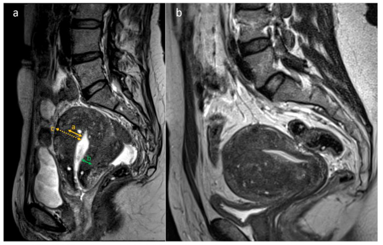
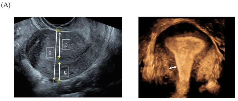
Our aim was to compare the inter-rater agreement about transvaginal ultrasonography (TVS) with magnetic resonance imaging (MRI) with regard to diagnosing adenomyosis and for assessing various predefined imaging features of adenomyosis, in the same set of women. The study cohort included 51 women, prospectively, consecutively recruited based on a clinical suspicion of adenomyosis. MRIs and TVS videoclips and 3D volumes were retrospectively assessed by four experienced radiologists and five experienced sonographers, respectively. Each rater subjectively evaluated the presence or absence of adenomyosis, as well as imaging features suggestive of adenomyosis. Fleiss kappa (κ) was used to reflect inter-rater agreement for categorical data, and the intraclass correlation coefficient (ICC) was used to reflect the reliability of quantitative data. Agreement between raters for diagnosing adenomyosis was higher for TVS than for MRI (κ = 0.42 vs. 0.28). MRI had a higher inter-rater agreement in assessing wall asymmetry, irregular junctional zone (JZ), and the presence of myometrial cysts, while TVU had a better agreement for assessing globular shape. MRI showed a moderate to good reliability for measuring the JZ (ICC = 0.57-0.82). For TVS, the JZ was unmeasurable in >50% of cases, and the remaining cases had low reliability (ICC = -0.31-0.08). We found that inter-rater agreement for diagnosing adenomyosis was higher for TVS than for MRI, despite the fact that MRI showed a higher inter-rater agreement in most specific features. Measurements of JZ in the coronal plane with 3D TVS were unreliable and thus unlikely to be useful for diagnosing adenomyosis.

摘要

我们的目的是比较同一组女性中,经阴道超声检查(TVS)与磁共振成像(MRI)在诊断子宫腺肌病以及评估子宫腺肌病各种预定义影像特征方面的评分者间一致性。研究队列包括51名女性,她们基于子宫腺肌病的临床疑似情况被前瞻性、连续招募。MRI以及TVS视频片段和三维容积分别由四名经验丰富的放射科医生和五名经验丰富的超声检查医师进行回顾性评估。每位评分者主观评估子宫腺肌病的有无以及提示子宫腺肌病的影像特征。Fleiss卡方(κ)用于反映分类数据的评分者间一致性,组内相关系数(ICC)用于反映定量数据的可靠性。TVS诊断子宫腺肌病时评分者间的一致性高于MRI(κ = 0.42对0.28)。MRI在评估肌壁不对称、交界区不规则(JZ)以及肌层囊肿的存在方面评分者间一致性更高,而TVS在评估球形方面一致性更好。MRI测量JZ显示出中度到良好的可靠性(ICC = 0.57 - 0.8)。对于TVS,超过50%的病例无法测量JZ,其余病例可靠性较低(ICC = -0.31 - 0.08)。我们发现,尽管MRI在大多数特定特征方面评分者间一致性更高,但TVS诊断子宫腺肌病时评分者间的一致性高于MRI。用三维TVS在冠状面测量JZ不可靠,因此不太可能用于诊断子宫腺肌病。

https://cdn.ncbi.nlm.nih.gov/pmc/blobs/f154/10341351/e4c319fb6276/diagnostics-13-02193-g002a.jpg
https://cdn.ncbi.nlm.nih.gov/pmc/blobs/f154/10341351/68d2cb472357/diagnostics-13-02193-g001.jpg
https://cdn.ncbi.nlm.nih.gov/pmc/blobs/f154/10341351/e4c319fb6276/diagnostics-13-02193-g002a.jpg
https://cdn.ncbi.nlm.nih.gov/pmc/blobs/f154/10341351/68d2cb472357/diagnostics-13-02193-g001.jpg
https://cdn.ncbi.nlm.nih.gov/pmc/blobs/f154/10341351/e4c319fb6276/diagnostics-13-02193-g002a.jpg

相似文献

1
Inter-Rater Agreement for Diagnosing Adenomyosis Using Magnetic Resonance Imaging and Transvaginal Ultrasonography.使用磁共振成像和经阴道超声诊断子宫腺肌病的评分者间一致性
Diagnostics (Basel). 2023 Jun 28;13(13):2193. doi: 10.3390/diagnostics13132193.
2
Inter-rater agreement in the diagnosis of adenomyosis by 2- and 3-dimensional transvaginal ultrasonography.二维和三维经阴道超声检查诊断子宫腺肌病的评分者间一致性
J Ultrasound Med. 2019 Mar;38(3):657-666. doi: 10.1002/jum.14735. Epub 2018 Sep 4.
3
Transvaginal sonographic features of diffuse adenomyosis in 18-30-year-old nulligravid women without endometriosis: association with symptoms.18至30岁未孕且无子宫内膜异位症的女性弥漫性子宫腺肌病的经阴道超声特征:与症状的关联
Ultrasound Obstet Gynecol. 2015 Dec;46(6):730-6. doi: 10.1002/uog.14834.
4
Adenomyosis: three-dimensional sonographic findings of the junctional zone and correlation with histology.子宫腺肌病:结合带的三维超声表现及其与组织学的相关性。
Ultrasound Obstet Gynecol. 2011 Apr;37(4):471-9. doi: 10.1002/uog.8900.
5
Three-dimensional ultrasound in diagnosis of adenomyosis: histologic correlation with ultrasound targeted biopsies of the uterus.三维超声在子宫腺肌病诊断中的应用:与超声靶向子宫活检的组织学相关性。
J Minim Invasive Gynecol. 2013 Nov-Dec;20(6):803-10. doi: 10.1016/j.jmig.2013.05.002.
6
Diagnosing adenomyosis with MRI: a prospective study revisiting the junctional zone thickness cutoff of 12 mm as a diagnostic marker.用 MRI 诊断子宫腺肌病:一项重新探讨交界带厚度 12 毫米作为诊断标志物的前瞻性研究。
Eur Radiol. 2019 Dec;29(12):6971-6981. doi: 10.1007/s00330-019-06308-3. Epub 2019 Jul 1.
7
Magnetic resonance imaging and transvaginal ultrasonography for the diagnosis of adenomyosis.磁共振成像和经阴道超声检查在子宫腺肌病诊断中的应用
Fertil Steril. 2001 Sep;76(3):588-94. doi: 10.1016/s0015-0282(01)01962-8.
8
The prediction of pouch of Douglas obliteration using offline analysis of the transvaginal ultrasound 'sliding sign' technique: inter- and intra-observer reproducibility.经阴道超声“滑动征”技术的离线分析预测道格拉斯窝消失:观察者间和观察者内的可重复性。
Hum Reprod. 2013 May;28(5):1237-46. doi: 10.1093/humrep/det044. Epub 2013 Mar 12.
9
Vaginal bromocriptine for treatment of adenomyosis: Impact on magnetic resonance imaging and transvaginal ultrasound.阴道用溴隐亭治疗子宫腺肌病:对磁共振成像和经阴道超声的影响
Eur J Obstet Gynecol Reprod Biol. 2020 Nov;254:38-43. doi: 10.1016/j.ejogrb.2020.08.040. Epub 2020 Aug 24.
10
Diagnostic Role of Transvaginal Sonography and Magnetic Resonance Imaging in Adenomyosis of the Uterus and its Correlation with Histopathology.经阴道超声检查和磁共振成像在子宫腺肌病中的诊断作用及其与组织病理学的相关性
J Midlife Health. 2023 Jan-Mar;14(1):34-41. doi: 10.4103/jmh.jmh_230_22. Epub 2023 Jul 7.

引用本文的文献

1
How Reproducible Are the Ultrasound Features of Adenomyosis Defined by the Revised MUSA Consensus?经修订的MUSA共识所定义的子宫腺肌病超声特征的可重复性如何?
J Clin Med. 2025 Jan 13;14(2):456. doi: 10.3390/jcm14020456.

本文引用的文献

1
Transvaginal ultrasound versus magnetic resonance imaging for diagnosing adenomyosis: A systematic review and head-to-head meta-analysis.经阴道超声与磁共振成像诊断子宫腺肌病的比较:系统评价和头对头荟萃分析。
Int J Gynaecol Obstet. 2023 May;161(2):397-405. doi: 10.1002/ijgo.14609. Epub 2022 Dec 11.
2
Consensus on revised definitions of Morphological Uterus Sonographic Assessment (MUSA) features of adenomyosis: results of modified Delphi procedure.关于子宫腺肌病形态学超声评估(MUSA)特征修订定义的共识:改良 Delphi 程序的结果。
Ultrasound Obstet Gynecol. 2022 Jul;60(1):118-131. doi: 10.1002/uog.24786.
3
Objective measures of adenomyosis on MRI and their diagnostic accuracy-a systematic review & meta-analysis.
MRI 对子宫腺肌病的客观测量及其诊断准确性的系统评价和荟萃分析。
Acta Obstet Gynecol Scand. 2021 Aug;100(8):1377-1391. doi: 10.1111/aogs.14139. Epub 2021 Apr 4.
4
Recommendations for MRI technique in the evaluation of pelvic endometriosis: consensus statement from the Society of Abdominal Radiology endometriosis disease-focused panel.盆腔子宫内膜异位症 MRI 技术评估的推荐意见:来自腹部放射学会子宫内膜异位症疾病重点专家组的共识声明。
Abdom Radiol (NY). 2020 Jun;45(6):1569-1586. doi: 10.1007/s00261-020-02483-w.
5
Noninvasive Diagnosis of Adenomyosis: A Structured Review and Meta-analysis of Diagnostic Accuracy in Imaging.影像学对子宫腺肌病的无创性诊断:系统评价和诊断准确性的 Meta 分析。
J Minim Invasive Gynecol. 2020 Feb;27(2):408-418.e3. doi: 10.1016/j.jmig.2019.11.001. Epub 2019 Nov 8.
6
Diagnosing adenomyosis with MRI: a prospective study revisiting the junctional zone thickness cutoff of 12 mm as a diagnostic marker.用 MRI 诊断子宫腺肌病:一项重新探讨交界带厚度 12 毫米作为诊断标志物的前瞻性研究。
Eur Radiol. 2019 Dec;29(12):6971-6981. doi: 10.1007/s00330-019-06308-3. Epub 2019 Jul 1.
7
The REDCap consortium: Building an international community of software platform partners.REDCap 联盟:构建软件平台合作伙伴的国际社区。
J Biomed Inform. 2019 Jul;95:103208. doi: 10.1016/j.jbi.2019.103208. Epub 2019 May 9.
8
Intra- and Inter-Rater Agreement Describing Myometrial Lesions Using Morphologic Uterus Sonographic Assessment: A Pilot Study.形态学子宫超声评估描述子宫肌瘤的组内和组间一致性:一项初步研究。
J Ultrasound Med. 2019 Oct;38(10):2673-2683. doi: 10.1002/jum.14971. Epub 2019 Feb 23.
9
A sonographic classification of adenomyosis: interobserver reproducibility in the evaluation of type and degree of the myometrial involvement.子宫腺肌病的超声分类:评估肌层受累类型和程度的观察者间可重复性。
Fertil Steril. 2018 Nov;110(6):1154-1161.e3. doi: 10.1016/j.fertnstert.2018.06.031.
10
Inter-rater agreement in the diagnosis of adenomyosis by 2- and 3-dimensional transvaginal ultrasonography.二维和三维经阴道超声检查诊断子宫腺肌病的评分者间一致性
J Ultrasound Med. 2019 Mar;38(3):657-666. doi: 10.1002/jum.14735. Epub 2018 Sep 4.