Jain Shivi, Kumar Kamlesh, Shukla Ram Chandra, Jain Madhu
Department of Radiodiagnosis and Imaging, Institute of Medical Sciences, Banaras Hindu University, Varanasi, Uttar Pradesh, India.
Department of Obstetrics and Gynaecology, Institute of Medical Sciences, Banaras Hindu University, Varanasi, Uttar Pradesh, India.
J Midlife Health. 2023 Jan-Mar;14(1):34-41. doi: 10.4103/jmh.jmh_230_22. Epub 2023 Jul 7.
The prevalence of adenomyosis of the uterus varies from 5% to 70%, and there is no clear consensus on its imaging diagnostic criteria. The objective of this study was to evaluate the role of transvaginal sonography (TVS), combined TVS and color Doppler (TVS-CD), and magnetic resonance imaging (MRI) in the diagnosis of adenomyosis.
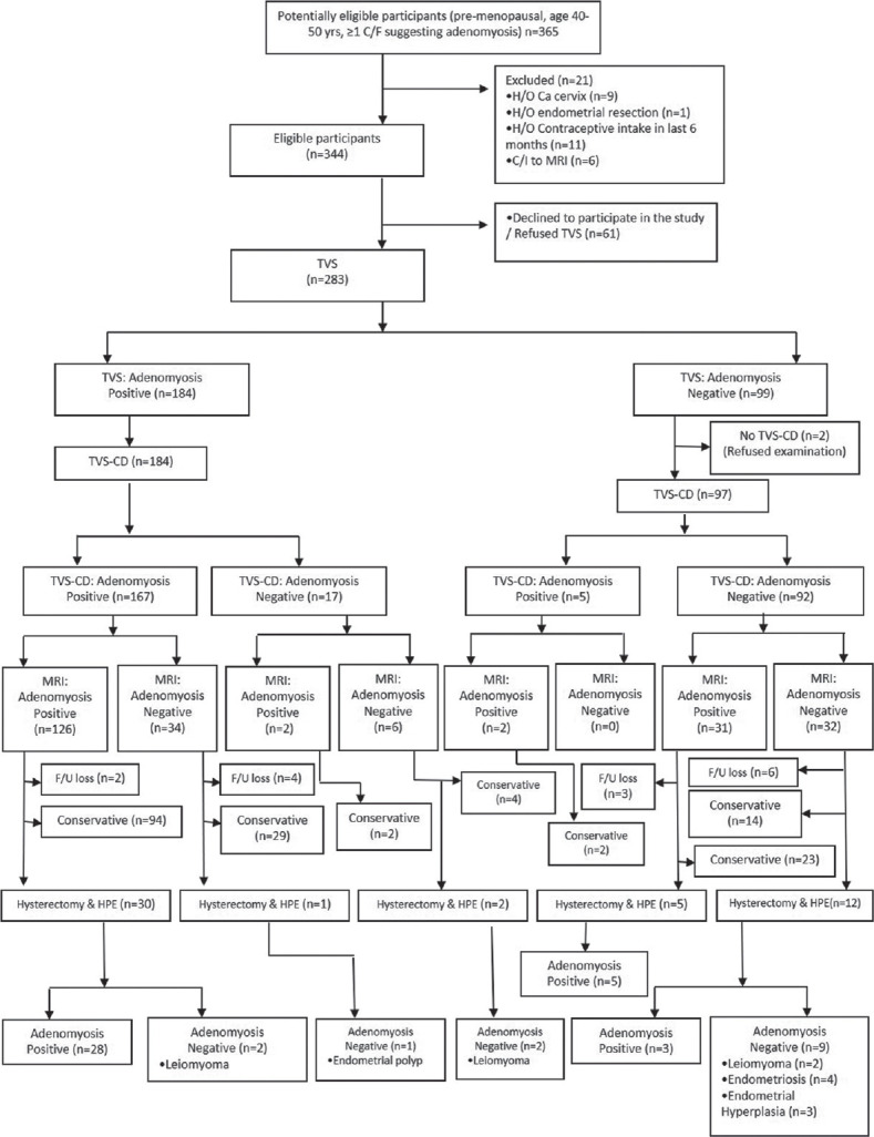
This was a tertiary care hospital-based prospective study, in which 365 clinically suspected cases of adenomyosis were enrolled. All three types of imaging (TVS, TVS-CD, and MRI) were done in 233/365 patients, followed by hysterectomy in 50. Imaging features were correlated with the histopathological examination (HPE), which was taken as the gold standard for the diagnosis. The diagnostic performance of each imaging modality was assessed.
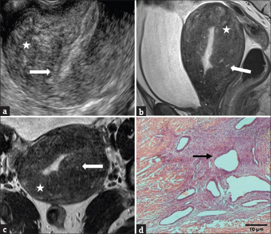
Among patients who underwent hysterectomy, 36/50 (72%) had adenomyosis on HPE, with or without associated benign gynecological abnormalities. Sensitivity, specificity, positive predictive value (PPV), negative PV (NPV), and diagnostic accuracy (DA) of MRI were higher than that of TVS-CD (91.67% vs. 77.78%, 85.71% vs. 78.57%, 94.29% vs. 90.32%, 80% vs. 57.89%, and 90% vs. 78%, respectively). TVS alone had lower diagnostic performance (specificity: 64.29%, PPV 84.85%, NPV 52.94%, and DA74%) than TVS-CD, but equal sensitivity (77.78%). Heterogeneous myometrium was the most sensitive (80.56%), while myometrial cyst was the most specific (92.86%) TVS feature. The maximum junctional zone thickness ≥12 mm was the most sensitive (97.22%), while the hyperintense myometrial focus was the most specific (100%) MRI feature.
TVS-CD should be used as an initial diagnostic imaging modality in clinically suspected cases of adenomyosis; however, MRI due to better diagnostic efficacy should be the imaging modality of choice before subjecting such patients to hysterectomy.
子宫腺肌病的患病率在5%至70%之间,其影像学诊断标准尚无明确共识。本研究的目的是评估经阴道超声(TVS)、TVS联合彩色多普勒(TVS-CD)及磁共振成像(MRI)在子宫腺肌病诊断中的作用。
这是一项基于三级医疗医院的前瞻性研究,纳入365例临床疑似子宫腺肌病的病例。233/365例患者接受了所有三种影像学检查(TVS、TVS-CD和MRI),其中50例随后接受了子宫切除术。将影像学特征与组织病理学检查(HPE)进行关联,HPE被视为诊断的金标准。评估了每种影像学检查方法的诊断性能。
在接受子宫切除术的患者中,36/50(72%)在HPE检查中患有子宫腺肌病,伴有或不伴有相关的良性妇科异常。MRI的敏感性、特异性、阳性预测值(PPV)、阴性预测值(NPV)和诊断准确性(DA)均高于TVS-CD(分别为91.67%对77.78%、85.71%对78.57%、94.29%对90.32%、80%对57.89%、90%对78%)。单独的TVS诊断性能低于TVS-CD(特异性:64.29%,PPV 84.85%,NPV 52.94%,DA 74%),但敏感性相同(77.78%)。肌层不均质是最敏感的TVS特征(80.56%),而肌层囊肿是最具特异性的TVS特征(92.86%)。结合带最大厚度≥12mm是最敏感的MRI特征(97.22%),而肌层高信号灶是最具特异性的MRI特征(100%)。
在临床疑似子宫腺肌病的病例中,TVS-CD应作为初始诊断影像学检查方法;然而,由于诊断效能更高,MRI应是此类患者接受子宫切除术之前的首选影像学检查方法。