Ma Zechen, Mulder Douwe Johannes, Gniadecki Robert, Cohen Tervaert Jan Willem, Osman Mohammed
Division of Rheumatology, Department of Medicine, University of Alberta, Edmonton, AB T6G 2R7, Canada.
Division of Vascular Medicine, Department of Internal Medicine, University Medical Centre Groningen, University of Groningen, 9700 RB Groningen, The Netherlands.
Diagnostics (Basel). 2023 Jun 28;13(13):2204. doi: 10.3390/diagnostics13132204.
Nailfolds of patients with systemic sclerosis (SSc) provide an opportunity to directly visualize microvascular remodeling in SSc. Nailfold video capillaroscopy (NVC) remains the gold standard for assessing nailfold capillaroscopy (NFC). However, access to NVC is limited by expense and expertise. This review aims to synthesize current research on other NFC devices compared to NVC.
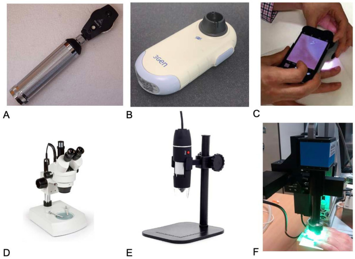
The literature search included the primary research of adult patients with SSc as defined by the 2013 ACR/EULAR criteria. Methods of assessing NFC included stereomicroscopy/wide-field microscopy, ophthalmoscopy, dermatoscopy, smartphone devices, and digital USB microscopy. Primary outcomes included both qualitative (normal vs. abnormal nailfolds, overall pattern recognition, presence/absence of giant capillaries, hemorrhages, and abnormal morphology) and quantitative (capillary density and dimension) measures.
The search yielded 471 studies, of which 9 were included. Five studies compared NVC to dermatoscopy, two compared it to widefield/stereomicroscopy, one to smartphone attachments, and one to USB microscopy. In dermatoscopy studies, NVC had a higher percentage of images that were interpretable (63-77% vs. 100%), classifiable (70% vs. 84%), or gradable (70% vs. 79.3%) across three studies. Dermatoscopy had a lower sensitivity (60.2% vs. 81.6%) and higher specificity (92.5% vs. 84.6%) compared to NVC. One stereomicroscopy study found a significant difference between methods in capillary density in limited cutaneous SSc, while another found correlations in all parameters between stereomicroscopy and NVC. One smartphone lens had good agreement with NVC on abnormal capillary morphology and density. USB microscopy was able to differentiate between SSc and healthy controls using mean capillary width but not by capillary density.
A dermatoscope may serve as a more portable and affordable screening tool to identify a normal "scleroderma pattern", and images that need further corroboration by NVC. NFC parameters reported are heterogenous and the standardization of these parameters is important, especially in non-gold-standard devices.
系统性硬化症(SSc)患者的甲襞为直接观察SSc中的微血管重塑提供了机会。甲襞视频毛细血管镜检查(NVC)仍然是评估甲襞毛细血管镜检查(NFC)的金标准。然而,NVC的使用受到费用和专业知识的限制。本综述旨在综合目前关于其他NFC设备与NVC相比的研究。
文献检索包括对符合2013年美国风湿病学会/欧洲抗风湿病联盟(ACR/EULAR)标准定义的成年SSc患者的初步研究。评估NFC的方法包括立体显微镜/广角显微镜、检眼镜检查、皮肤镜检查、智能手机设备和数字USB显微镜检查。主要结果包括定性(正常与异常甲襞、整体模式识别、有无巨型毛细血管、出血和异常形态)和定量(毛细血管密度和尺寸)指标。
检索共获得471项研究,其中9项被纳入。5项研究将NVC与皮肤镜检查进行比较,2项将其与广角/立体显微镜检查进行比较,1项与智能手机附件进行比较,1项与USB显微镜检查进行比较。在皮肤镜检查研究中,在三项研究中,NVC具有可解释图像(63 - 77%对100%)、可分类图像(70%对84%)或可分级图像(70%对79.3%)的比例更高。与NVC相比,皮肤镜检查的敏感性较低(60.2%对81.6%),特异性较高(92.5%对84.6%)。一项立体显微镜检查研究发现,在局限性皮肤型SSc中,两种方法在毛细血管密度方面存在显著差异,而另一项研究发现立体显微镜检查与NVC在所有参数上均具有相关性。一种智能手机镜头在异常毛细血管形态和密度方面与NVC具有良好的一致性。USB显微镜检查能够使用平均毛细血管宽度区分SSc和健康对照,但不能通过毛细血管密度进行区分。
皮肤镜可作为一种更便携且经济的筛查工具,用于识别正常的“硬皮病模式”以及需要NVC进一步证实的图像。所报告的NFC参数存在异质性,这些参数的标准化很重要,尤其是在非金标准设备中。