Institut de Recerca de l'Hospital de la Santa Creu i Sant Pau, Institut d'Investigació Biomèdica (IIB) Sant Pau, 08041 Barcelona, Spain.
Department of Biochemistry and Molecular Biology, Universitat Autònoma de Barcelona (UAB), 08025 Barcelona, Spain.
Int J Mol Sci. 2023 Jul 6;24(13):11153. doi: 10.3390/ijms241311153.
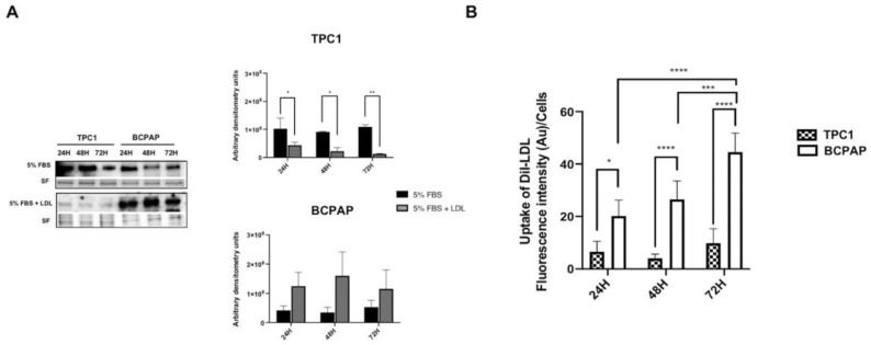
We previously described the role of low-density lipoprotein (LDL) in aggressiveness in papillary thyroid cancer (PTC). Moreover, the MAPK signaling pathway in the presence of BRAF V600E mutation is associated with more aggressive PTC. Although the link between MAPK cascade and LDL receptor (LDLR) expression has been previously described, it is unknown whether LDL can potentiate the adverse effects of PTC through it. We aimed to investigate whether the presence of LDL might accelerate the oncogenic processes through MAPK pathway in presence or absence of BRAF V600E in two thyroid cell lines: TPC1 and BCPAP (wild-type and BRAF V600E, respectively). LDLR, PI3K-AKT and RAS/RAF/MAPK (MEK)/ERK were analyzed via Western blot; cell proliferation was measured via MTT assay, cell migration was studied through wound-healing assay and LDL uptake was analyzed by fluorometric and confocal analysis. TPC1 demonstrated a time-specific downregulation of the LDLR, while BCPAP resulted in a receptor deregulation after LDL exposition. LDL uptake was increased in BCPAP over-time, as well as cell proliferation (20% higher) in comparison to TPC1. Both cell lines differed in migration pattern with a wound closure of 83.5 ± 9.7% after LDL coculture in TPC1, while a loss in the adhesion capacity was detected in BCPAP. The siRNA knockdown of LDLR in LDL-treated BCPAP cells resulted in a p-ERK expression downregulation and cell proliferation modulation, demonstrating a link between LDLR and MAPK pathway. The modulation of BRAF-V600E using vemurafenib-impaired LDLR expression decreased cellular proliferation. Our results suggest that LDLR regulation is cell line-specific, regulating the RAS/RAF/MAPK (MEK)/ERK pathway in the LDL-signaling cascade and where BRAF V600E can play a critical role. In conclusion, targeting LDLR and this downstream signaling cascade, could be a new therapeutic strategy for PTC with more aggressive behavior, especially in those harboring BRAF V600E.
我们之前描述了低密度脂蛋白(LDL)在甲状腺乳头状癌(PTC)侵袭性中的作用。此外,存在 BRAF V600E 突变的 MAPK 信号通路与侵袭性更强的 PTC 相关。尽管之前已经描述了 MAPK 级联与 LDL 受体(LDLR)表达之间的联系,但尚不清楚 LDL 是否可以通过它增强 PTC 的不良影响。我们旨在研究在两种甲状腺细胞系(TPC1 和 BCPAP[野生型和 BRAF V600E])中,LDL 是否存在通过 MAPK 通路加速致癌过程,无论是否存在 BRAF V600E。通过 Western blot 分析 LDLR、PI3K-AKT 和 RAS/RAF/MAPK(MEK)/ERK;通过 MTT 测定法测量细胞增殖,通过划痕愈合测定法研究细胞迁移,通过荧光和共聚焦分析分析 LDL 摄取。TPC1 表现出 LDLR 的时间特异性下调,而 BCPAP 在 LDL 暴露后导致受体失调。随着时间的推移,BCPAP 中的 LDL 摄取增加,细胞增殖(比 TPC1 高 20%)增加。与 TPC1 相比,两种细胞系在迁移模式上存在差异,LDL 共培养后 TPC1 的伤口闭合率为 83.5±9.7%,而 BCPAP 则检测到粘附能力丧失。LDL 处理的 BCPAP 细胞中 LDLR 的 siRNA 敲低导致 p-ERK 表达下调和细胞增殖调节,表明 LDLR 与 MAPK 通路之间存在联系。使用 vemurafenib 调节 BRAF-V600E 降低了 LDLR 表达,从而减少了细胞增殖。我们的结果表明,LDLR 调节是细胞系特异性的,调节 LDL 信号级联中的 RAS/RAF/MAPK(MEK)/ERK 通路,而 BRAF V600E 可以在此发挥关键作用。总之,针对 LDLR 和这个下游信号级联,可能是治疗侵袭性更强的 PTC 的一种新的治疗策略,特别是在那些携带 BRAF V600E 的患者中。