Department of Rare Disorders, Norwegian Centre of Expertise for Neurodevelopmental Disorders and Hypersomnias (NevSom), Oslo University Hospital, Ullevål, Oslo, Norway.
Division of Mental Health and Addiction, NORMENT Centre, University of Oslo and Oslo University Hospital, Oslo, Norway.
Sleep. 2023 Nov 8;46(11). doi: 10.1093/sleep/zsad173.
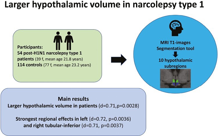
Narcolepsy type 1 (NT1) is a neurological sleep disorder. Postmortem studies have shown 75%-90% loss of the 50 000-70 000 hypocretin-producing neurons and 64%-94% increase in the 64 000-120 000 histaminergic neurons and conflicting indications of gliosis in the hypothalamus of NT1 patients. The aim of this study was to compare MRI-based volumes of the hypothalamus in patients with NT1 and controls in vivo.
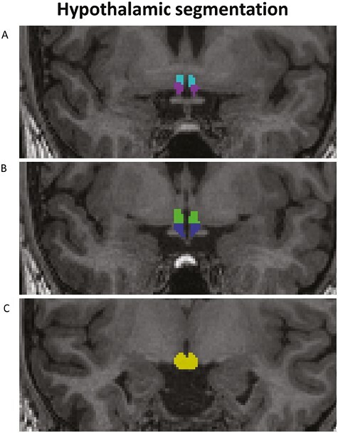
We used a segmentation tool based on deep learning included in Freesurfer and computed the volume of the whole hypothalamus, left/right part of the hypothalamus, and 10 hypothalamic subregions. We included 54 patients with post-H1N1 NT1 (39 females, mean age 21.8 ± 11.0 years) and 114 controls (77 females, mean age 23.2 ± 9.0 years). Group differences were tested with general linear models using permutation testing in Permutation Analysis of Linear Models and evaluated after 10 000 permutations, yielding two-tailed P-values. Furthermore, a stepwise Bonferroni correction was performed after dividing hypothalamus into smaller regions.
The analysis revealed larger volume for patients compared to controls for the whole hypothalamus (Cohen's d = 0.71, p = 0.0028) and for the left (d = 0.70, p = 0.0037) and right part of the hypothalamus (d = 0.65, p = 0.0075) and left (d = 0.72, p = 0.0036) and right tubular-inferior (d = 0.71, p = 0.0037) hypothalamic subregions.
In conclusion, patients with post-H1N1 NT1 showed significantly larger hypothalamic volume than controls, in particular in the tubular-inferior subregions which could reflect several processes as previous studies have indicated neuroinflammation, gliosis, and changes in the numbers of different cell types.
发作性睡病 1 型(NT1)是一种神经睡眠障碍。尸检研究表明,NT1 患者下丘脑的下丘脑泌素能神经元减少了 75%-90%,而组胺能神经元增加了 64%-94%,并且存在神经胶质增生的矛盾迹象。本研究旨在比较 NT1 患者和对照组的下丘脑磁共振成像(MRI)体积。
我们使用了 Freesurfer 中包含的基于深度学习的分割工具,计算了整个下丘脑、下丘脑左右部分以及 10 个下丘脑亚区的体积。我们纳入了 54 例甲型 H1N1 后 NT1 患者(39 名女性,平均年龄 21.8±11.0 岁)和 114 名对照组(77 名女性,平均年龄 23.2±9.0 岁)。使用置换分析线性模型中的置换检验,通过一般线性模型测试组间差异,并在进行 10000 次置换后评估双侧 P 值。此外,在将下丘脑划分为较小区域后,我们进行了逐步 Bonferroni 校正。
分析显示,与对照组相比,患者的整个下丘脑(Cohen's d=0.71,p=0.0028)、左侧(d=0.70,p=0.0037)和右侧(d=0.65,p=0.0075)以及左侧(d=0.72,p=0.0036)和右侧管状下(d=0.71,p=0.0037)下丘脑亚区的体积更大。
总之,甲型 H1N1 后发作性睡病 1 型患者的下丘脑体积明显大于对照组,特别是在管状下亚区,这可能反映了几项过程,如先前的研究表明的神经炎症、神经胶质增生以及不同细胞类型数量的变化。