Centre for Medical Image Computing, Department of Medical Physics and Biomedical Engineering, University College, London, UK.
Dementia Research Centre, Department of Neurodegenerative Disease, UCL Queen Square Institute of Neurology, University College, London, UK.
Neuroimage. 2020 Dec;223:117287. doi: 10.1016/j.neuroimage.2020.117287. Epub 2020 Aug 25.
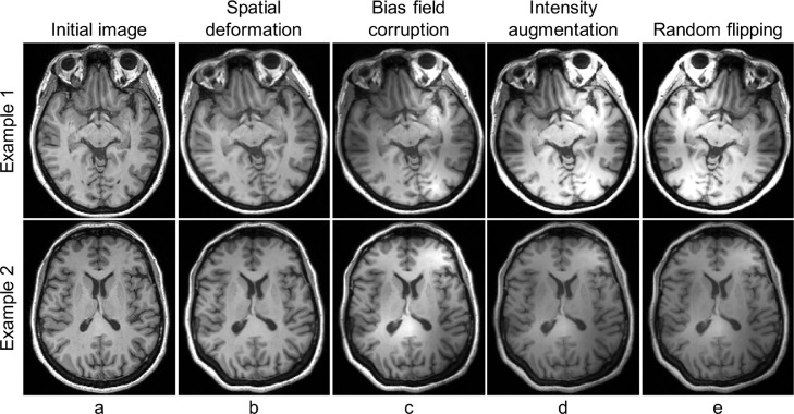
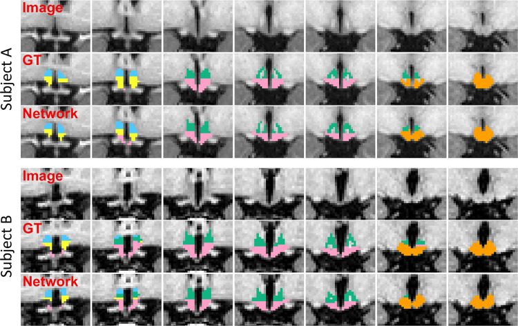
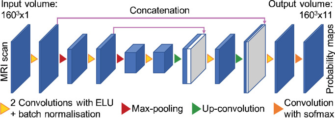
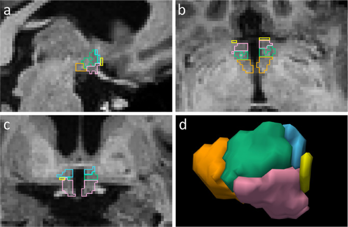
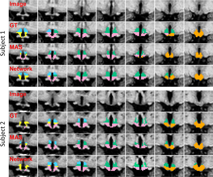
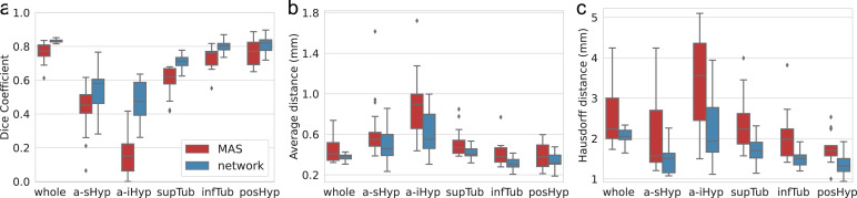
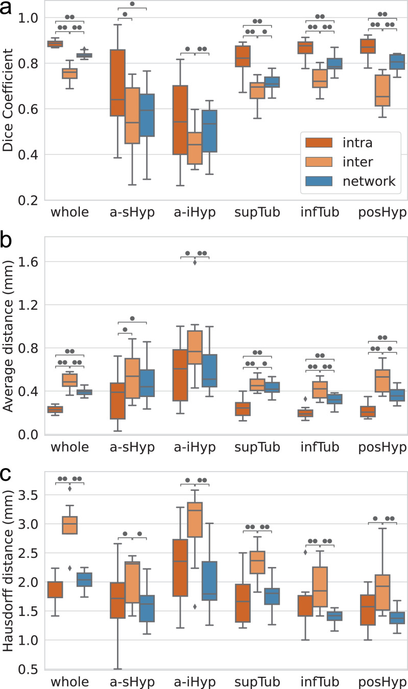
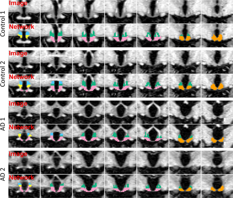
Despite the crucial role of the hypothalamus in the regulation of the human body, neuroimaging studies of this structure and its nuclei are scarce. Such scarcity partially stems from the lack of automated segmentation tools, since manual delineation suffers from scalability and reproducibility issues. Due to the small size of the hypothalamus and the lack of image contrast in its vicinity, automated segmentation is difficult and has been long neglected by widespread neuroimaging packages like FreeSurfer or FSL. Nonetheless, recent advances in deep machine learning are enabling us to tackle difficult segmentation problems with high accuracy. In this paper we present a fully automated tool based on a deep convolutional neural network, for the segmentation of the whole hypothalamus and its subregions from T1-weighted MRI scans. We use aggressive data augmentation in order to make the model robust to T1-weighted MR scans from a wide array of different sources, without any need for preprocessing. We rigorously assess the performance of the presented tool through extensive analyses, including: inter- and intra-rater variability experiments between human observers; comparison of our tool with manual segmentation; comparison with an automated method based on multi-atlas segmentation; assessment of robustness by quality control analysis of a larger, heterogeneous dataset (ADNI); and indirect evaluation with a volumetric study performed on ADNI. The presented model outperforms multi-atlas segmentation scores as well as inter-rater accuracy level, and approaches intra-rater precision. Our method does not require any preprocessing and runs in less than a second on a GPU, and approximately 10 seconds on a CPU. The source code as well as the trained model are publicly available at https://github.com/BBillot/hypothalamus_seg, and will also be distributed with FreeSurfer.
尽管下丘脑在人体调节中起着至关重要的作用,但针对该结构及其核的神经影像学研究却很少。这种稀缺性部分源于缺乏自动化分割工具,因为手动描绘存在可扩展性和可重复性问题。由于下丘脑体积较小,且其附近的图像对比度不足,因此自动化分割较为困难,并且长期以来一直被像 FreeSurfer 或 FSL 这样的广泛使用的神经影像学套件所忽视。尽管如此,深度学习的最新进展使我们能够高精度地解决困难的分割问题。在本文中,我们提出了一种完全基于深度卷积神经网络的自动化工具,用于从 T1 加权 MRI 扫描中分割整个下丘脑及其子区域。我们使用激进的数据增强方法,使模型能够对来自各种不同来源的 T1 加权 MRI 扫描具有鲁棒性,而无需任何预处理。我们通过广泛的分析,包括:人类观察者之间的组内和组间变异性实验;与手动分割的比较;与基于多图谱分割的自动方法的比较;通过对更大、更异构数据集(ADNI)的质量控制分析来评估稳健性;以及在 ADNI 上进行的容积研究的间接评估,来严格评估所提出工具的性能。所提出的模型在多图谱分割评分以及组内变异性实验中表现优于观察者间的准确性水平,并且接近观察者内的精确性。我们的方法不需要任何预处理,在 GPU 上的运行时间不到一秒,在 CPU 上的运行时间约为 10 秒。源代码以及训练好的模型可在 https://github.com/BBillot/hypothalamus_seg 上获取,并且也将随 FreeSurfer 一起发布。