Luo Christine T, Yee Jennifer
The Ohio State University, Department of Emergency Medicine, Columbus OH.
J Educ Teach Emerg Med. 2020 Jan 15;5(1):S26-S52. doi: 10.21980/J8T938. eCollection 2020 Jan.
The aim of this simulation case is to educate senior medical students, resident physicians, and advanced practice providers on the recognition, diagnosis, and management of spinal epidural abscesses. This scenario is most applicable to the emergency medicine setting but can be applied to the outpatient office or urgent care settings.
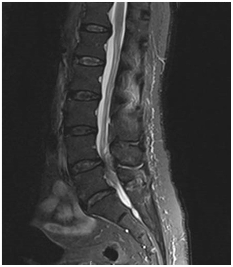
Spinal epidural abscess is an infection leading to an epidural collection of purulent material. This uncommon condition is estimated to occur less than 12 times per 100,000 hospital admissions.1,2 However, this infection can lead to devastating neurological sequelae via cord compression, spinal vascular interruption, and inflammatory etiologies;3,4 thus, prompt diagnosis is essential. Unfortunately, spinal epidural abscesses may be difficult to identify clinically due to variable clinical presentations. The goal of this scenario is to increase awareness of this critical diagnosis.Detailed history-taking to identify risk factors will aid in the recognition of spinal epidural abscesses. Many of the risk factors are related to increased infectious risk from hematogenous spread, iatrogenic inoculation, or direct extension.1 Individuals with conditions including intravenous (IV) drug use, alcohol abuse, diabetes, human immunodeficiency virus (HIV), cancer, hepatic disease, renal disease, and other immunocompromising conditions are at increased risk of developing epidural abscesses.1 Primary infectious sources include dental abscesses, endocarditis, vertebral osteomyelitis, and soft tissue infections. Spinal procedures including spinal surgeries, paraspinal injections, and placement of epidural catheters or stimulators can also predispose to infection.2,4Classic symptoms for spinal epidural abscesses include fever, back pain and neurological changes.1,5 Back pain is the most frequent presenting symptom, occurring about 70%-90% of the time.1 However, fever is the least frequent presenting symptom4 and neurological findings only occur in about one-third of cases.2 Neurological symptoms include motor weakness, sensory changes, urinary retention, overflow urinary incontinence, bowel dysfunction, hyperreflexia, radicular pain, spinal shock or cauda equina syndrome.1,4Laboratory findings may include systemic leukocytosis and elevated inflammatory markers. Whereas leukocytosis is estimated to be present in two-thirds of cases,2 Davis, et al. showed that with the concurrent presence of a risk factor, an elevated erythrocyte sedimentation rate (ESR) had 100% sensitivity and 67% specificity for spinal epidural abscesses.5Magnetic resonance imaging (MRI) with gadolinium contrast is the preferred imaging modality for diagnosing spinal epidural abscesses. Computed tomography (CT) with myelography can be considered if MRI is contraindicated.1 Given that abscesses may be multifocal, further spinal imaging beyond a single spinal segment should be considered during evaluation. Lumbar puncture is not recommended due to risk of iatrogenic infectious spread.Treatment of epidural abscesses includes obtaining blood cultures and prompt antibiotic administration with early surgical evaluation to determine if operative intervention is warranted. is the most common microbial cause, contributing to about two-thirds of cases.3,4 Other microbial causes include coagulase-negative (ie, ), , gram-negative bacilli (ie, and ), and less commonly, anaerobic bacteria, fungi, mycobacteria and parasites.1,2 Empiric antibiotic treatments generally include vancomycin and a third- or fourth- generation cephalosporin.2,4This simulation session will highlight the importance of recognizing and aggressively treating this uncommon but potentially devastating condition.
After this simulation case, learners will be able to diagnose and manage patients with spinal epidural abscesses. Specifically, learners will be able to:Obtain a detailed history, including past infectious, surgical, procedural and social history to evaluate for epidural abscess risk factors. Describe clinical signs and symptoms of spinal epidural abscesses and understand that initial clinical presentations can be variable.Perform a focused neurological exam including evaluation of motor, sensory, reflexes, and rectal tone.Order appropriate laboratory testing and imaging modalities for spinal epidural abscess diagnosis, including a post-void bladder residual volume.Select appropriate antibiotics for empiric treatment of spinal epidural abscess depending on patient presentation.Disposition the patient to appropriate inpatient care.
The authors conducted this simulation case with a standardized patient. We encourage inclusion of a standardized patient versus a mannequin to provide appropriate motor and sensory exams. For those without a standardized patient program, the authors suggest utilizing a faculty member as the patient. Regardless of individual used, it is strongly recommended that facilitators rehearse the case with the individual in the patient role ahead of time in order to ensure that their performance reflects an accurate neurologic exam. A debriefing session and small-group discussion followed the simulation to review the clinical presentation, diagnosis, management, and treatment of spinal epidural abscesses. This case can also be adapted as an oral boards case.
Residents were provided a survey at the completion of the debriefing session to rate different aspects of the simulation, as well as to provide qualitative feedback on the scenario. This survey is specific to our institution's simulation center.
While qualitative feedback from the residents was positive, it was viewed as a straightforward case. Our initial presenting symptom was difficulty ambulating with a fever at home, if asked. The residents appreciated performing a neurologic exam on a standardized patient versus attempting this on a mannequin.Our simulation center's feedback form is based on the Center of Medical Simulation's Debriefing Assessment for Simulation in Healthcare (DASH) Student Version Short Form with the inclusion of required qualitative feedback if an element was scored less than a 6 or 7. This session received all 7 scores (extremely effective/outstanding) other than one 5 score for the element assessing if the instructor set the stage for an engaging learning experience. The learner's feedback for this 5 score was "kinda went right into the case which was ok." Our form also includes an area for general feedback about the case at the end. Comments included "Great sim. Expert case writing," "Fun case and learned a lot," and "Great case! Appreciated feedback on consulting and the difficult consultant situation."
This is a cost-effective method for reviewing epidural abscess. We chose a chief complaint and history that was slightly atypical from "classic" presentations, but not so esoteric that the residents felt cheated at the end of the scenario. When using a standardized patient in a scenario that may involve a sensitive physical exam, we review with learners and the standardized patient what expectations are during the pre-brief session. For example, residents may say, "we would like to check to see if rectal tone is intact," and then the standardized patient would verbalize back the expected physical exam findings.
Medical simulation, spinal epidural abscess, spinal cord compression, infectious disease.
本模拟病例的目的是对高年级医学生、住院医师和高级执业医师进行脊髓硬膜外脓肿的识别、诊断和管理方面的培训。此场景最适用于急诊医学环境,但也可应用于门诊办公室或紧急护理环境。
脊髓硬膜外脓肿是一种导致硬膜外脓性物质聚集的感染。这种罕见疾病估计每10万例住院患者中发生不到12次。然而,这种感染可通过脊髓压迫、脊髓血管中断和炎症病因导致严重的神经后遗症;因此,及时诊断至关重要。不幸的是,由于临床表现多样,脊髓硬膜外脓肿在临床上可能难以识别。本场景的目的是提高对这一关键诊断的认识。详细询问病史以确定危险因素将有助于识别脊髓硬膜外脓肿。许多危险因素与血行播散、医源性接种或直接蔓延导致的感染风险增加有关。患有静脉注射毒品使用、酗酒、糖尿病、人类免疫缺陷病毒(HIV)、癌症、肝病、肾病和其他免疫功能低下疾病的个体发生硬膜外脓肿的风险增加。主要感染源包括牙脓肿、心内膜炎、椎体骨髓炎和软组织感染。包括脊柱手术、椎旁注射以及硬膜外导管或刺激器置入在内的脊柱操作也易引发感染。脊髓硬膜外脓肿的典型症状包括发热、背痛和神经功能改变。背痛是最常见的症状,约70%-90%的病例会出现。然而,发热是最不常见的症状,神经学表现仅在约三分之一的病例中出现。神经症状包括运动无力、感觉改变、尿潴留、充溢性尿失禁、肠道功能障碍、反射亢进神经根性疼痛、脊髓休克或马尾综合征。实验室检查结果可能包括全身白细胞增多和炎症标志物升高。虽然估计三分之二的病例会出现白细胞增多,但戴维斯等人表明,在存在危险因素的情况下,红细胞沉降率(ESR)升高对脊髓硬膜外脓肿的敏感性为100%,特异性为67%。钆增强磁共振成像(MRI)是诊断脊髓硬膜外脓肿的首选影像学检查方法。如果MRI禁忌,可考虑计算机断层扫描(CT)脊髓造影。鉴于脓肿可能是多灶性的,在评估过程中应考虑对单个脊髓节段以外的脊髓进行进一步成像检查。由于存在医源性感染传播的风险,不建议进行腰椎穿刺。硬膜外脓肿的治疗包括进行血培养并及时给予抗生素,同时尽早进行手术评估以确定是否需要手术干预。金黄色葡萄球菌是最常见的微生物病因,约占三分之二的病例。其他微生物病因包括凝固酶阴性葡萄球菌(即表皮葡萄球菌)、链球菌、革兰氏阴性杆菌(即大肠杆菌和肺炎克雷伯菌),较少见的还有厌氧菌、真菌、分枝杆菌和寄生虫。经验性抗生素治疗通常包括万古霉素和第三代或第四代头孢菌素。本次模拟课程将强调识别和积极治疗这种罕见但可能具有毁灭性的疾病的重要性。
完成本模拟病例后,学习者将能够诊断和管理脊髓硬膜外脓肿患者。具体而言,学习者将能够:获取详细病史,包括既往感染、手术、操作和社会史,以评估硬膜外脓肿的危险因素。描述脊髓硬膜外脓肿的临床体征和症状,并了解初始临床表现可能各不相同。进行重点神经系统检查,包括评估运动、感觉、反射和直肠张力。为脊髓硬膜外脓肿的诊断开具适当的实验室检查和影像学检查项目,包括排尿后膀胱残余尿量检查。根据患者表现选择适当的抗生素进行脊髓硬膜外脓肿的经验性治疗。将患者安排到适当的住院护理。
作者使用标准化患者进行了本模拟病例。我们鼓励纳入标准化患者而非人体模型,以便进行适当的运动和感觉检查。对于没有标准化患者项目的机构,作者建议利用教员扮演患者。无论使用何种个体,强烈建议教员提前与扮演患者角色的个体排练病例,以确保其表现反映准确的神经系统检查。模拟结束后进行了总结会议和小组讨论,以回顾脊髓硬膜外脓肿的临床表现、诊断、管理和治疗。此病例也可改编为口试病例。
在总结会议结束时,向住院医师提供了一份调查问卷,以对模拟的不同方面进行评分,并就该场景提供定性反馈。此调查问卷特定于我们机构的模拟中心。
虽然住院医师的定性反馈是积极的,但他们认为这是一个简单直接的病例。我们最初设定的症状是在家中行走困难并伴有发热(如果被问到)。住院医师们赞赏在标准化患者身上进行神经系统检查,而不是在人体模型上尝试。我们模拟中心的反馈表基于医疗模拟中心的医疗保健模拟总结评估(DASH)学生版简表,并在某个要素得分低于6或7时纳入所需的定性反馈。除了评估教员是否为引人入胜学习体验搭建舞台这一要素得分为5分之外,本次课程的所有要素得分均为7分(极其有效/出色)。对于这个5分的要素,学习者的反馈是“有点直接进入病例了,不过还行”。我们的表格在最后还包括一个关于该病例的总体反馈区域。评论包括“很棒的模拟。专业的病例编写”,“有趣的病例,学到了很多”,以及“很棒的病例!感谢关于会诊和困难会诊情况的反馈”。
这是一种复习硬膜外脓肿的经济有效方法。我们选择了一个主诉和病史,与“经典”表现略有不同,但又不至于深奥到让住院医师在场景结束时感到受骗。在可能涉及敏感体格检查的场景中使用标准化患者时,我们在预演环节与学习者和标准化患者一起回顾期望的内容。例如,住院医师可能会说,“我们想检查一下直肠张力是否正常”,然后标准化患者会说出预期的体格检查结果。
医学模拟、脊髓硬膜外脓肿、脊髓压迫、传染病。