Hunt Derek Jc, McLendon Kevin, Wiggins Matthew
Merit Health Wesley, Department of Emergency Medicine, Hattiesburg, MS.
J Educ Teach Emerg Med. 2020 Apr 19;6(2):V8-V12. doi: 10.21980/J8J644. eCollection 2021 Apr.
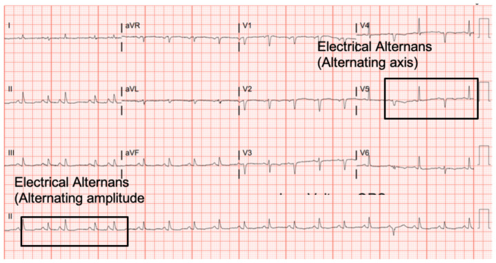
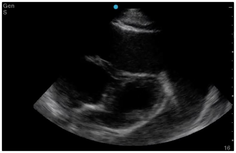
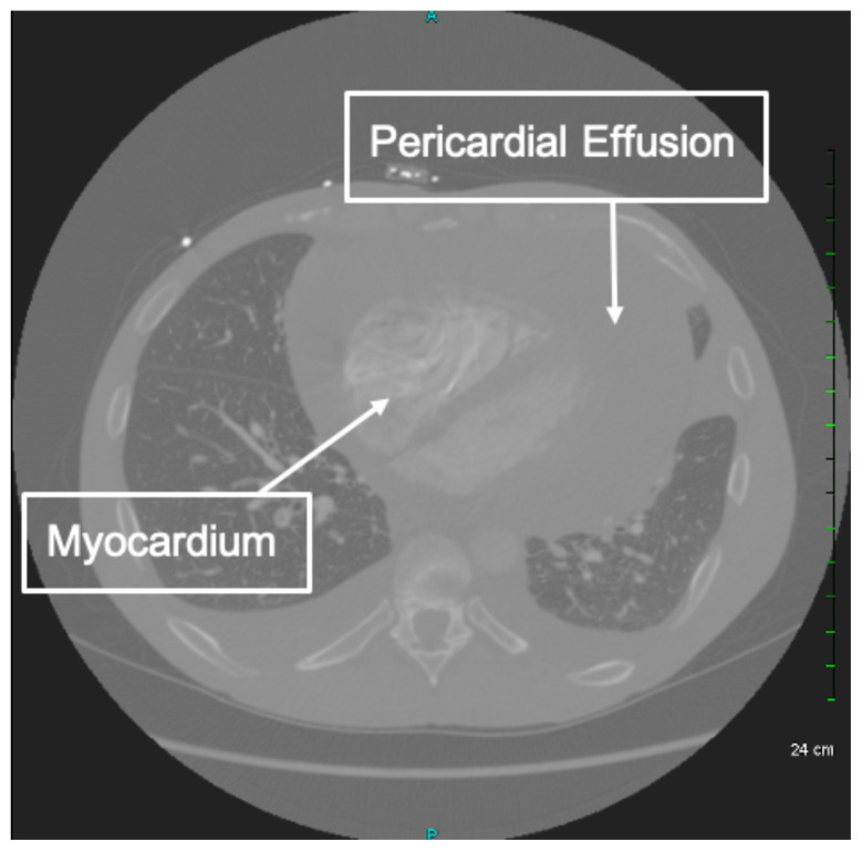
Cardiac tamponade is a rare and life-threatening condition that can be caused by trauma and various medical conditions. Failure to recognize and repair it quickly could lead to significant morbidity or even death. This case demonstrates the electrocardiogram (ECG) findings of low voltage QRS and electrical alternans in cardiac tamponade. It also highlights the classic ultrasound (US) findings of pericardial effusion and right ventricular collapse during diastole in cardiac tamponade. Classic physical exam findings of cardiac tamponade include Beck's Triad (jugular venous distention, hypotension, and muffled heart sounds) and pulsus paradoxus. This patient only had jugular venous distention and pulsus paradoxus. The case is centered on a 52-year-old male who presented with shortness of breath, wheezing, and a productive cough with streaks of blood. A CT chest was performed which revealed a large pericardial effusion, right upper lobe lung mass, and bilateral pulmonary emboli. A bedside transthoracic echocardiogram was then performed which confirmed the large effusion as well as right ventricular collapse during diastole. Cardiothoracic surgery and interventional cardiology were consulted and both agreed to take the patient to the cardiac catheterization lab for percutaneous drainage of the effusion. Pericardiocentesis was performed and 1.7 liters of serosanguinous fluid was removed and a drain was left in place. He recovered well from the procedure and had an uneventful admission. After reviewing this case, learners should be able to recognize the diagnostic features and various causes of pericardial effusion and cardiac tamponade.
Electrocardiography, echocardiography, cardiac tamponade.
心脏压塞是一种罕见且危及生命的病症,可由创伤和各种疾病引起。未能迅速识别并修复它可能导致严重的发病甚至死亡。本病例展示了心脏压塞时低电压QRS波和电交替的心电图(ECG)表现。它还突出了心脏压塞时心包积液和舒张期右心室塌陷的典型超声(US)表现。心脏压塞的典型体格检查发现包括贝克三联征(颈静脉怒张、低血压和心音低钝)和奇脉。该患者仅有颈静脉怒张和奇脉。该病例围绕一名52岁男性展开,他出现呼吸急促、喘息和伴有血丝的咳痰。进行了胸部CT检查,结果显示大量心包积液、右上叶肺肿块和双侧肺栓塞。随后进行了床边经胸超声心动图检查,证实了大量积液以及舒张期右心室塌陷。咨询了心胸外科和介入心脏病学专家,他们都同意将患者送往心脏导管实验室进行积液的经皮引流。进行了心包穿刺术,抽出了1.7升浆液性血性液体,并留置了引流管。他术后恢复良好,住院过程顺利。在回顾这个病例后,学习者应该能够识别心包积液和心脏压塞的诊断特征及各种病因。
心电图、超声心动图、心脏压塞。