• 文献检索
  • 文档翻译
  • 深度研究
  • 学术资讯
  • Suppr Zotero 插件Zotero 插件
  • 邀请有礼
  • 套餐&价格
  • 历史记录
应用&插件
Suppr Zotero 插件Zotero 插件浏览器插件Mac 客户端Windows 客户端微信小程序
定价
高级版会员购买积分包购买API积分包
服务
文献检索文档翻译深度研究API 文档MCP 服务
关于我们
关于 Suppr公司介绍联系我们用户协议隐私条款
关注我们

Suppr 超能文献

核心技术专利:CN118964589B侵权必究
粤ICP备2023148730 号-1Suppr @ 2026

文献检索

告别复杂PubMed语法,用中文像聊天一样搜索,搜遍4000万医学文献。AI智能推荐,让科研检索更轻松。

立即免费搜索

文件翻译

保留排版,准确专业,支持PDF/Word/PPT等文件格式,支持 12+语言互译。

免费翻译文档

深度研究

AI帮你快速写综述,25分钟生成高质量综述,智能提取关键信息,辅助科研写作。

立即免费体验

一名40岁不吸烟患者原发性肺腺癌表现为心脏压塞

Primary Lung Adenocarcinoma Presenting as Cardiac Tamponade in a 40-Year-Old Non-Smoker.

作者信息

Dessalegn Noah, Felux Kelsee, Seid Ekram, Mohammed Amir

机构信息

Internal Medicine, Wellstar Atlanta Medical Center, Atlanta, USA.

Internal Medicine, Ross University School of Medicine, Miramar, USA.

出版信息

Cureus. 2022 Jan 26;14(1):e21631. doi: 10.7759/cureus.21631. eCollection 2022 Jan.

DOI:10.7759/cureus.21631
PMID:35228977
原文链接:https://pmc.ncbi.nlm.nih.gov/articles/PMC8877732/
Abstract

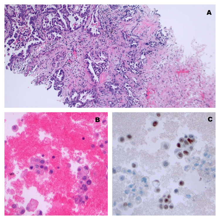
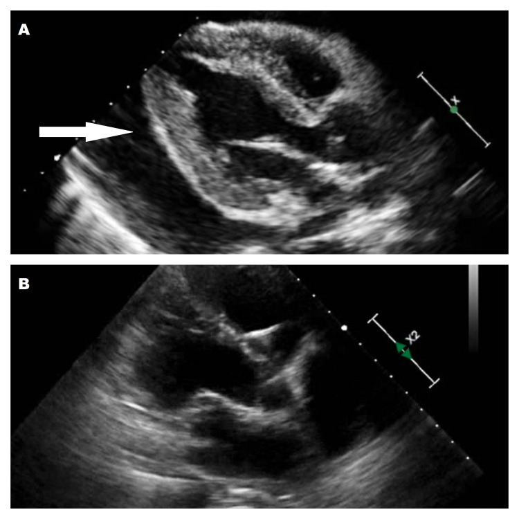
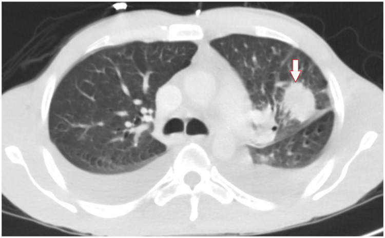
Lung cancer is the number one cause of cancer-death in the world with the majority of cases directly attributable to smoking. The diagnosis is mostly made following evaluation for either an incidental lung nodule or respiratory signs and symptoms such as cough and hemoptysis. This is a review of a young never-smoker who presented predominantly with gastrointestinal symptoms, which is an uncommon initial presentation of lung cancer associated with malignant pericardial effusion. A 40-year-old male without a history of smoking presented with epigastric pain associated with nausea and vomiting. He denied significant cardio-respiratory or systemic symptoms. Physical examination was unremarkable besides tachycardia of 111 beats per minute, blood pressure of 108/65 mmHg, and mild generalized direct abdominal tenderness. EKG showed electrical alternans. CXR demonstrated a prominent cardiac silhouette leading to evaluation with echocardiography, which revealed a large pericardial effusion and signs of cardiac tamponade. 1200 ml of serosanguinous fluid was removed by pericardiocentesis with significant clinical improvement. The basic workup of infectious and immunologic causes was negative, which prompted a contrasted CT scan of the chest. This revealed a left upper lobe mass measuring 3.6 x 2.8 cm without mediastinal or hilar lymphadenopathy. CT-guided biopsy was performed and was consistent with pulmonary adenocarcinoma but was negative for molecular drivers and programmed cell death ligand 1 (PD-L1). Pericardial fluid cytology also confirmed the presence of malignant cells. The patient complained of mild dyspnea and chest pain before discharge which led to a repeat echocardiogram and identification of a recurrent large pericardial effusion. Cardiothoracic surgery consultation was obtained, and the patient underwent subxiphoid pericardial window placement. Learning points from this case report include: First, non-smoking-related lung cancer is still among the top ten causes of cancer death in the US. It should remain in the differential diagnosis of patients presenting with pertinent signs and symptoms, even in non-smokers. Secondly, malignancy, most importantly primary lung cancer, is a common cause of a large symptomatic pericardial effusion in patients who have a non-revealing basic workup. In such patients, a detailed evaluation for undetected underlying malignancy is important. Thirdly, colchicine and non-steroidal anti-inflammatory drugs are commonly used for the treatment of painful malignant pericardial effusion; however, there is a lack of data to support this practice. Finally, pre-discharge screening echocardiography in patients with new or recurring cardiorespiratory symptoms following initial pericardiocentesis could be important because recurrent large pericardial effusion is a common and potentially fatal complication of malignant pericardial effusion.

https://cdn.ncbi.nlm.nih.gov/pmc/blobs/848e/8877732/f9885918744c/cureus-0014-00000021631-i04.jpg
https://cdn.ncbi.nlm.nih.gov/pmc/blobs/848e/8877732/9832f4e36a21/cureus-0014-00000021631-i01.jpg
https://cdn.ncbi.nlm.nih.gov/pmc/blobs/848e/8877732/9278b314c37b/cureus-0014-00000021631-i02.jpg
https://cdn.ncbi.nlm.nih.gov/pmc/blobs/848e/8877732/3429b6dc6c93/cureus-0014-00000021631-i03.jpg
https://cdn.ncbi.nlm.nih.gov/pmc/blobs/848e/8877732/f9885918744c/cureus-0014-00000021631-i04.jpg
摘要

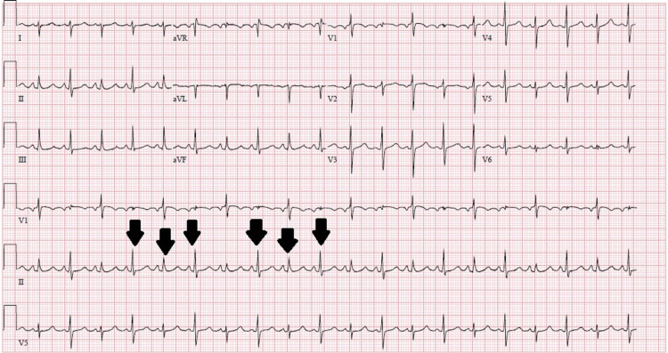
肺癌是全球癌症死亡的首要原因,大多数病例直接归因于吸烟。诊断大多是在对偶然发现的肺结节或咳嗽、咯血等呼吸道症状和体征进行评估后做出的。本文回顾了一名主要表现为胃肠道症状的年轻不吸烟者,这是肺癌合并恶性心包积液的一种不常见的初始表现。一名40岁无吸烟史男性,出现上腹部疼痛并伴有恶心、呕吐。他否认有明显的心肺或全身症状。体格检查除每分钟111次的心动过速、血压108/65 mmHg和轻度全身性腹部直接压痛外无异常。心电图显示电交替。胸部X线显示心脏轮廓突出,遂行超声心动图检查,结果显示大量心包积液及心脏压塞征象。通过心包穿刺抽出1200毫升浆液性血性液体,临床症状明显改善。感染和免疫病因的基本检查均为阴性,这促使进行胸部增强CT扫描。结果显示左肺上叶有一大小为3.6×2.8 cm的肿块,无纵隔或肺门淋巴结肿大。进行了CT引导下活检,结果符合肺腺癌,但分子驱动因素和程序性细胞死亡配体1(PD-L1)检测为阴性。心包积液细胞学检查也证实存在恶性细胞。患者出院前诉轻度呼吸困难和胸痛,这导致复查超声心动图并发现复发性大量心包积液。获得心胸外科会诊意见后,患者接受了剑突下心包开窗术。本病例报告的经验教训包括:第一,与吸烟无关的肺癌仍是美国癌症死亡的十大原因之一。即使在不吸烟者中,对于出现相关症状和体征的患者,仍应将其列入鉴别诊断。第二,恶性肿瘤,最重要的是原发性肺癌,是基础检查无异常的患者出现有症状的大量心包积液的常见原因。对于此类患者,详细评估未被发现的潜在恶性肿瘤很重要。第三,秋水仙碱和非甾体抗炎药常用于治疗疼痛性恶性心包积液;然而,缺乏支持这种做法的数据。最后,对于首次心包穿刺后出现新的或复发性心肺症状的患者,出院前进行超声心动图筛查可能很重要,因为复发性大量心包积液是恶性心包积液常见且可能致命的并发症。

相似文献

1
Primary Lung Adenocarcinoma Presenting as Cardiac Tamponade in a 40-Year-Old Non-Smoker.一名40岁不吸烟患者原发性肺腺癌表现为心脏压塞
Cureus. 2022 Jan 26;14(1):e21631. doi: 10.7759/cureus.21631. eCollection 2022 Jan.
2
Diagnosis of malignant pericarditis: a single centre experience.恶性心包炎的诊断:单中心经验。
Kardiol Pol. 2012;70(11):1147-53.
3
Malignant pericardial effusion with cardiac tamponade in ovarian adenocarcinoma.卵巢腺癌合并恶性心包积液并心脏压塞。
Arch Gynecol Obstet. 2009 Oct;280(4):675-8. doi: 10.1007/s00404-009-0976-5. Epub 2009 Feb 19.
4
Anaplastic Lymphoma Kinase-Positive Primary Lung Adenocarcinoma Presenting With Pericardial Effusion and Tamponade in a COVID-19 Patient: A Case Report.一名COVID-19患者出现心包积液和心脏压塞的间变性淋巴瘤激酶阳性原发性肺腺癌:病例报告
Cureus. 2021 Oct 29;13(10):e19127. doi: 10.7759/cureus.19127. eCollection 2021 Oct.
5
Intrapericardial cisplatin administration after pericardiocentesis in patients with lung adenocarcinoma and malignant cardiac tamponade.肺腺癌合并恶性心包填塞患者心包穿刺术后心包腔内注射顺铂
Hellenic J Cardiol. 2005 Sep-Oct;46(5):324-9.
6
Hemorrhagic Pericardial Effusion Leading to Cardiac Tamponade: A Rare Initial Presentation of Adenocarcinoma of the Lung.出血性心包积液导致心脏压塞:肺癌的一种罕见首发表现
Cureus. 2020 Nov 10;12(11):e11411. doi: 10.7759/cureus.11411.
7
Cardiac Tamponade in Down's Syndrome Associated With Hypothyroidism: An Uncommon Presentation.唐氏综合征合并甲状腺功能减退症患者的心脏压塞:一种罕见的表现。
Cureus. 2024 Apr 25;16(4):e59023. doi: 10.7759/cureus.59023. eCollection 2024 Apr.
8
The role of early contrast-enhanced chest computed tomography in the aetiological diagnosis of patients presenting with cardiac tamponade or large pericardial effusion.早期对比增强胸部计算机断层扫描在心脏压塞或大量心包积液患者病因诊断中的作用。
Eur Heart J Cardiovasc Imaging. 2016 Apr;17(4):421-8. doi: 10.1093/ehjci/jev225. Epub 2015 Sep 15.
9
A Case Report of Cardiac Tamponade.心脏压塞一例报告
J Educ Teach Emerg Med. 2020 Apr 19;6(2):V8-V12. doi: 10.21980/J8J644. eCollection 2021 Apr.
10
Mediastinal effusion due to pericardiocentesis with cardiac tamponade: a case report.心包穿刺导致的心包积液压迫:病例报告。
BMC Anesthesiol. 2021 Jun 16;21(1):172. doi: 10.1186/s12871-021-01385-8.

引用本文的文献

1
Incidental finding of lung adenocarcinoma following routine bullectomy and pleurodesis on a young patient: a case report.一名年轻患者在常规肺大疱切除术和胸膜固定术后偶然发现肺腺癌:病例报告
JRSM Open. 2025 May 8;16(5):20542704241304218. doi: 10.1177/20542704241304218. eCollection 2025 May.
2
Misleading Chest Pain: Acute Coronary Syndrome or Lung Adenocarcinoma With Malignant Pericardial Effusion.误导性胸痛:急性冠状动脉综合征还是伴有恶性心包积液的肺腺癌
Cureus. 2024 Jul 29;16(7):e65624. doi: 10.7759/cureus.65624. eCollection 2024 Jul.
3
Lung Adenocarcinoma Presenting as Early Cardiac Tamponade: A Case Report.

本文引用的文献

1
Global Cancer Statistics 2020: GLOBOCAN Estimates of Incidence and Mortality Worldwide for 36 Cancers in 185 Countries.《全球癌症统计数据 2020:全球 185 个国家和地区 36 种癌症的发病率和死亡率估计》。
CA Cancer J Clin. 2021 May;71(3):209-249. doi: 10.3322/caac.21660. Epub 2021 Feb 4.
2
Cancer Statistics, 2021.癌症统计数据,2021.
CA Cancer J Clin. 2021 Jan;71(1):7-33. doi: 10.3322/caac.21654. Epub 2021 Jan 12.
3
Pericardial diseases in patients with cancer: contemporary prevalence, management and outcomes.癌症患者的心包疾病:当代患病率、处理方法和结局。
以早期心脏压塞为表现的肺腺癌:一例报告
Case Rep Oncol. 2024 Jul 25;17(1):779-787. doi: 10.1159/000540183. eCollection 2024 Jan-Dec.
4
A Case of Primary Lung Adenocarcinoma With Two Uncommon Presentations: Neurological Paraneoplastic Syndrome and Pericardial Effusion.一例具有两种罕见表现的原发性肺腺癌:神经副肿瘤综合征和心包积液
Cureus. 2023 Oct 26;15(10):e47753. doi: 10.7759/cureus.47753. eCollection 2023 Oct.
Heart. 2020 Apr;106(8):569-574. doi: 10.1136/heartjnl-2019-315852. Epub 2020 Jan 24.
4
Systematic review of percutaneous interventions for malignant pericardial effusion.恶性心包积液经皮介入治疗的系统评价
Heart. 2015 Oct;101(20):1619-26. doi: 10.1136/heartjnl-2015-307907. Epub 2015 Jul 15.
5
Lung cancer in never smokers--a different disease.从不吸烟者患肺癌——一种不同的疾病。
Nat Rev Cancer. 2007 Oct;7(10):778-90. doi: 10.1038/nrc2190.
6
Lung cancer in never smokers: a review.从不吸烟者的肺癌:综述
J Clin Oncol. 2007 Feb 10;25(5):561-70. doi: 10.1200/JCO.2006.06.8015.
7
Large symptomatic pericardial effusion as the presentation of unrecognized cancer: a study in 173 consecutive patients undergoing pericardiocentesis.大量有症状的心包积液作为未被识别癌症的表现:对173例连续接受心包穿刺术患者的研究
Medicine (Baltimore). 2006 Jan;85(1):49-53. doi: 10.1097/01.md.0000199556.69588.8e.
8
Malignant effusions.恶性胸腔积液
CA Cancer J Clin. 1991 May-Jun;41(3):166-79. doi: 10.3322/canjclin.41.3.166.