• 文献检索
  • 文档翻译
  • 深度研究
  • 学术资讯
  • Suppr Zotero 插件Zotero 插件
  • 邀请有礼
  • 套餐&价格
  • 历史记录
应用&插件
Suppr Zotero 插件Zotero 插件浏览器插件Mac 客户端Windows 客户端微信小程序
定价
高级版会员购买积分包购买API积分包
服务
文献检索文档翻译深度研究API 文档MCP 服务
关于我们
关于 Suppr公司介绍联系我们用户协议隐私条款
关注我们

Suppr 超能文献

核心技术专利:CN118964589B侵权必究
粤ICP备2023148730 号-1Suppr @ 2026

文献检索

告别复杂PubMed语法,用中文像聊天一样搜索,搜遍4000万医学文献。AI智能推荐,让科研检索更轻松。

立即免费搜索

文件翻译

保留排版,准确专业,支持PDF/Word/PPT等文件格式,支持 12+语言互译。

免费翻译文档

深度研究

AI帮你快速写综述,25分钟生成高质量综述,智能提取关键信息,辅助科研写作。

立即免费体验

3至4个月大人类胚胎颞下颌关节髁突的单细胞RNA测序分析

Single-cell RNA sequencing analysis of the temporomandibular joint condyle in 3 and 4-month-old human embryos.

作者信息

Zhu Qianqi, Tan Miaoying, Wang Chengniu, Chen Yufei, Wang Chenfei, Zhang Junqi, Gu Yijun, Guo Yuqi, Han Jianpeng, Li Lei, Jiang Rongrong, Fan Xudong, Xie Huimin, Wang Liang, Gu Zhifeng, Liu Dong, Shi Jianwu, Feng Xingmei

机构信息

Department of Stomatology, Affiliated Hospital of Nantong University, Medical School of, Nantong University, Nantong, 226001, China.

Institute of Reproductive Medicine, Medical School of Nantong University, Nantong, 226001, China.

出版信息

Cell Biosci. 2023 Jul 19;13(1):130. doi: 10.1186/s13578-023-01069-5.

DOI:10.1186/s13578-023-01069-5
PMID:37468984
原文链接:https://pmc.ncbi.nlm.nih.gov/articles/PMC10357633/
Abstract

BACKGROUND

The temporomandibular joint (TMJ) is a complex joint consisting of the condyle, the temporal articular surface, and the articular disc. Functions such as mastication, swallowing and articulation are accomplished by the movements of the TMJ. To date, the TMJ has been studied more extensively, but the types of TMJ cells, their differentiation, and their interrelationship during growth and development are still unclear and the study of the TMJ is limited. The aim of this study was to establish a molecular cellular atlas of the human embryonic temporomandibular joint condyle (TMJC) by single-cell RNA sequencing, which will contribute to understanding and solving clinical problems.

RESULTS

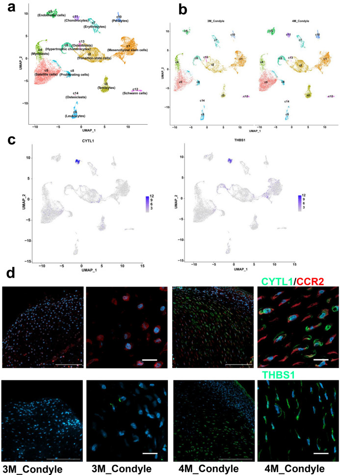
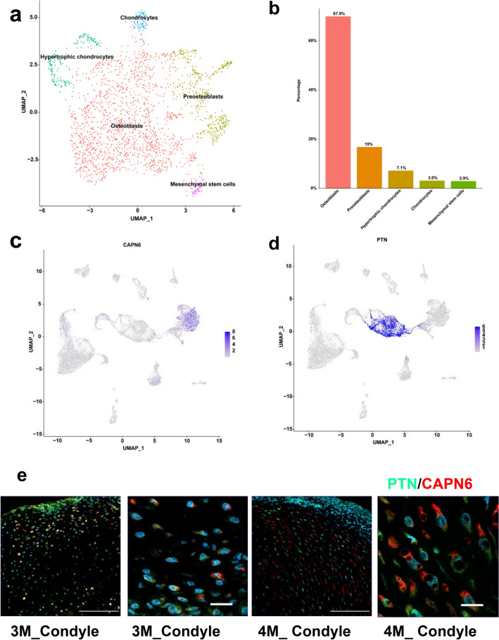
Human embryos at 3 and 4 months of age are an important stage of TMJC development. We performed a comprehensive transcriptome analysis of TMJC tissue from human embryos at 3 and 4 months of age using single-cell RNA sequencing. A total of 16,624 cells were captured and the gene expression profiles of 15 cell clusters in human embryonic TMJC were determined, including 14 known cell types and one previously unknown cell type, "transition state cells (TSCs)". Immunofluorescence assays confirmed that TSCs are not the same cell cluster as mesenchymal stem cells (MSCs). Pseudotime trajectory and RNA velocity analysis revealed that MSCs transformed into TSCs, which further differentiated into osteoblasts, hypertrophic chondrocytes and tenocytes. In addition, chondrocytes (CYTL1 + THBS1) from secondary cartilage were detected only in 4-month-old human embryonic TMJC.

CONCLUSIONS

Our study provides an atlas of differentiation stages of human embryonic TMJC tissue cells, which will contribute to an in-depth understanding of the pathophysiology of the TMJC tissue repair process and ultimately help to solve clinical problems.

摘要

背景

颞下颌关节(TMJ)是一个复杂的关节,由髁突、颞骨关节面和关节盘组成。咀嚼、吞咽和发音等功能通过颞下颌关节的运动来完成。迄今为止,对颞下颌关节的研究较为广泛,但颞下颌关节细胞的类型、它们的分化以及在生长发育过程中的相互关系仍不明确,对颞下颌关节的研究有限。本研究的目的是通过单细胞RNA测序建立人类胚胎颞下颌关节髁突(TMJC)的分子细胞图谱,这将有助于理解和解决临床问题。

结果

3个月和4个月大的人类胚胎是TMJC发育的重要阶段。我们使用单细胞RNA测序对3个月和4个月大的人类胚胎TMJC组织进行了全面的转录组分析。共捕获了16624个细胞,确定了人类胚胎TMJC中15个细胞簇的基因表达谱,包括14种已知细胞类型和一种以前未知的细胞类型,即“过渡态细胞(TSCs)”。免疫荧光分析证实TSCs与间充质干细胞(MSCs)不是同一细胞簇。伪时间轨迹和RNA速度分析显示,MSCs转化为TSCs,TSCs进一步分化为成骨细胞、肥大软骨细胞和肌腱细胞。此外,仅在4个月大的人类胚胎TMJC中检测到来自继发性软骨的软骨细胞(CYTL1+THBS1)。

结论

我们的研究提供了人类胚胎TMJC组织细胞分化阶段的图谱,这将有助于深入了解TMJC组织修复过程的病理生理学,并最终有助于解决临床问题。

https://cdn.ncbi.nlm.nih.gov/pmc/blobs/e182/10357633/db64c1838aac/13578_2023_1069_Fig6_HTML.jpg
https://cdn.ncbi.nlm.nih.gov/pmc/blobs/e182/10357633/f74b740e7d3c/13578_2023_1069_Fig1_HTML.jpg
https://cdn.ncbi.nlm.nih.gov/pmc/blobs/e182/10357633/fc51a960fb9c/13578_2023_1069_Fig2_HTML.jpg
https://cdn.ncbi.nlm.nih.gov/pmc/blobs/e182/10357633/188dcef4cb7e/13578_2023_1069_Fig3_HTML.jpg
https://cdn.ncbi.nlm.nih.gov/pmc/blobs/e182/10357633/a7d659ab61cc/13578_2023_1069_Fig4_HTML.jpg
https://cdn.ncbi.nlm.nih.gov/pmc/blobs/e182/10357633/087c5b019f2d/13578_2023_1069_Fig5_HTML.jpg
https://cdn.ncbi.nlm.nih.gov/pmc/blobs/e182/10357633/db64c1838aac/13578_2023_1069_Fig6_HTML.jpg

相似文献

1
Single-cell RNA sequencing analysis of the temporomandibular joint condyle in 3 and 4-month-old human embryos.3至4个月大人类胚胎颞下颌关节髁突的单细胞RNA测序分析
Cell Biosci. 2023 Jul 19;13(1):130. doi: 10.1186/s13578-023-01069-5.
2
Comparison of knee joint and temporomandibular joint development in pig embryos.猪胚胎膝关节与颞下颌关节发育的比较。
Anim Biotechnol. 2024 Nov;35(1):2337760. doi: 10.1080/10495398.2024.2337760. Epub 2024 Apr 24.
3
Development of the temporomandibular joint in miniature pig embryos.小型猪胚胎颞下颌关节的发育。
J Morphol. 2022 Jan;283(1):134-143. doi: 10.1002/jmor.21432. Epub 2021 Nov 27.
4
Expression of chondromodulin-1 in the temporomandibular joint condylar cartilage and disc.软骨调节素-1 在颞下颌关节髁状突软骨和盘的表达。
J Oral Pathol Med. 2010 Apr;39(4):356-60. doi: 10.1111/j.1600-0714.2009.00831.x. Epub 2009 Nov 5.
5
Development process of traumatic heterotopic ossification of the temporomandibular joint in mice.小鼠颞下颌关节创伤性异位骨化的发展过程。
J Craniomaxillofac Surg. 2019 Jul;47(7):1155-1161. doi: 10.1016/j.jcms.2018.11.026. Epub 2018 Dec 3.
6
Mesenchymal Stromal Cell Transplantation Induces Regeneration of Large and Full-Thickness Cartilage Defect of the Temporomandibular Joint.间质基质细胞移植诱导颞下颌关节大而全层软骨缺损的再生。
Cartilage. 2021 Dec;13(1_suppl):1814S-1821S. doi: 10.1177/1947603520926711. Epub 2020 Jun 4.
7
A comparison of human umbilical cord matrix stem cells and temporomandibular joint condylar chondrocytes for tissue engineering temporomandibular joint condylar cartilage.用于组织工程颞下颌关节髁突软骨的人脐带基质干细胞与颞下颌关节髁突软骨细胞的比较
Tissue Eng. 2007 Aug;13(8):2003-10. doi: 10.1089/ten.2006.0150.
8
The Roles of Indian Hedgehog Signaling in TMJ Formation.Indian Hedgehog 信号通路在 TMJ 形成中的作用。
Int J Mol Sci. 2019 Dec 13;20(24):6300. doi: 10.3390/ijms20246300.
9
Enhanced Circadian Clock in MSCs-Based Cytotherapy Ameliorates Age-Related Temporomandibular Joint Condyle Degeneration.基于间充质干细胞的细胞治疗增强生物钟可改善与年龄相关的颞下颌关节髁突退行性变。
Int J Mol Sci. 2021 Sep 30;22(19):10632. doi: 10.3390/ijms221910632.
10
Observing the development of the temporomandibular joint in embryonic and post-natal mice using various staining methods.使用各种染色方法观察胚胎期和出生后小鼠颞下颌关节的发育情况。
Exp Ther Med. 2016 Feb;11(2):481-489. doi: 10.3892/etm.2015.2937. Epub 2015 Dec 16.

引用本文的文献

1
Spatial Transcriptomics Unravel the Tissue Complexity of Oral Pathogenesis.空间转录组学揭示口腔发病机制的组织复杂性。
J Dent Res. 2024 Dec;103(13):1331-1339. doi: 10.1177/00220345241271934. Epub 2024 Oct 9.
2
Single‑cell RNA sequencing analysis of human embryos from the late Carnegie to fetal development.从卡内基晚期到胎儿发育阶段的人类胚胎单细胞RNA测序分析
Cell Biosci. 2024 Sep 12;14(1):118. doi: 10.1186/s13578-024-01302-9.

本文引用的文献

1
Taxonomy of fibroblasts and progenitors in the synovial joint at single-cell resolution.单细胞分辨率解析滑膜关节中的成纤维细胞和祖细胞分类。
Ann Rheum Dis. 2023 Mar;82(3):428-437. doi: 10.1136/ard-2021-221682. Epub 2022 Nov 22.
2
Single-Cell RNA Sequencing Reveals Transcriptional Changes in the Cartilage of Subchondral Insufficiency Fracture of the Knee.单细胞RNA测序揭示了膝关节软骨下骨不全骨折软骨的转录变化。
J Inflamm Res. 2022 Nov 5;15:6105-6112. doi: 10.2147/JIR.S385648. eCollection 2022.
3
Application of BMP in Bone Tissue Engineering.
骨形态发生蛋白在骨组织工程中的应用。
Front Bioeng Biotechnol. 2022 Mar 31;10:810880. doi: 10.3389/fbioe.2022.810880. eCollection 2022.
4
Overexpression of transcription factor FoxA2 in the developing skeleton causes an enlargement of the cartilage hypertrophic zone, but it does not trigger ectopic differentiation in immature chondrocytes.转录因子 FoxA2 在发育中的骨骼中的过度表达导致软骨肥大区的扩大,但它不会引发未成熟软骨细胞的异位分化。
Bone. 2022 Jul;160:116418. doi: 10.1016/j.bone.2022.116418. Epub 2022 Apr 6.
5
Modulation of miR-204 Expression during Chondrogenesis.在软骨发生过程中 miR-204 的表达调控。
Int J Mol Sci. 2022 Feb 15;23(4):2130. doi: 10.3390/ijms23042130.
6
ERα/β/DMP1 axis promotes trans-differentiation of chondrocytes to bone cells through GSK-3β/β-catenin pathway.ERα/β/DMP1 轴通过 GSK-3β/β-连环蛋白通路促进软骨细胞向成骨细胞的转分化。
J Anat. 2022 Jun;240(6):1152-1161. doi: 10.1111/joa.13612. Epub 2022 Jan 26.
7
Targeting the IL-6-Yap-Snail signalling axis in synovial fibroblasts ameliorates inflammatory arthritis.靶向滑膜成纤维细胞中的 IL-6-Yap-Snail 信号轴可改善炎症性关节炎。
Ann Rheum Dis. 2022 Feb;81(2):214-224. doi: 10.1136/annrheumdis-2021-220875. Epub 2021 Nov 29.
8
STAT3 is critical for skeletal development and bone homeostasis by regulating osteogenesis.STAT3 在骨骼发育和骨内稳态中起着关键作用,通过调节成骨作用。
Nat Commun. 2021 Nov 25;12(1):6891. doi: 10.1038/s41467-021-27273-w.
9
CNC-bZIP protein NFE2L1 regulates osteoclast differentiation in antioxidant-dependent and independent manners.CNC-bZIP蛋白NFE2L1以抗氧化剂依赖和非依赖的方式调节破骨细胞分化。
Redox Biol. 2021 Nov 6;48:102180. doi: 10.1016/j.redox.2021.102180.
10
LncRNA NEAT1 controls the lineage fates of BMSCs during skeletal aging by impairing mitochondrial function and pluripotency maintenance.长链非编码 RNA NEAT1 通过损害骨髓间充质干细胞的线粒体功能和多能性维持来控制其在骨骼衰老过程中的谱系命运。
Cell Death Differ. 2022 Feb;29(2):351-365. doi: 10.1038/s41418-021-00858-0. Epub 2021 Sep 8.