Adanyeguh Isaac M, Joers James M, Deelchand Dinesh K, Hutter Diane H, Eberly Lynn E, Guo Bin, Iltis Isabelle, Bushara Khalaf O, Henry Pierre-Gilles, Lenglet Christophe
Center for Magnetic Resonance Research and Department of Radiology, University of Minnesota Medical School, Minneapolis, MN 55455, USA.
Division of Biostatistics, School of Public Health, University of Minnesota, Minneapolis, MN 55455, USA.
Brain Commun. 2023 Jul 6;5(4):fcad196. doi: 10.1093/braincomms/fcad196. eCollection 2023.
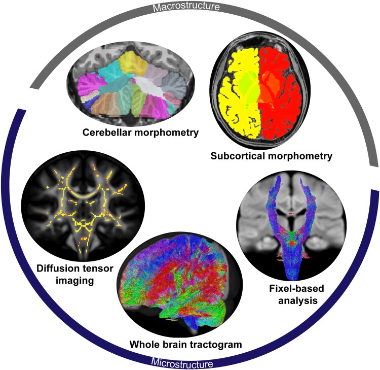
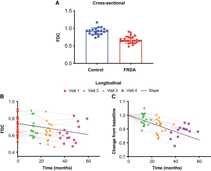
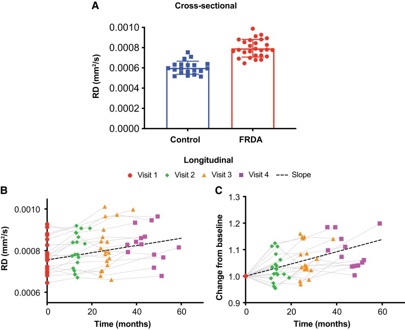
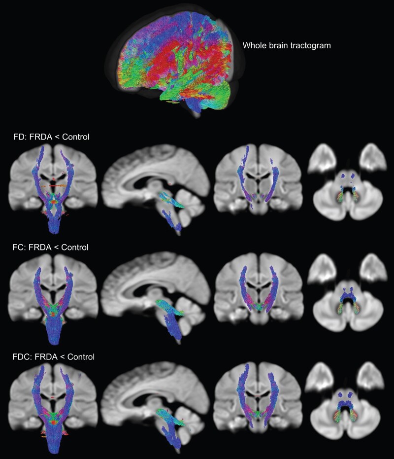
Friedreich ataxia is a progressive neurodegenerative disorder characterized by cerebellar and spinal atrophy. However, studies to elucidate the longitudinal progression of the pathology in the brain are somewhat inconsistent and limited, especially for early-stage Friedreich ataxia. Using a multimodal neuroimaging protocol, combined with advanced analysis methods, we sought to identify macrostructural and microstructural alterations in the brain of patients with early-stage Friedreich ataxia to better understand its distribution patterns and progression. We enrolled 28 patients with Friedreich ataxia and 20 age- and gender-matched controls. Longitudinal clinical and imaging data were collected in the patients at baseline, 12, 24 and 36 months. Macrostructural differences were observed in patients with Friedreich ataxia, compared to controls, including lower volume of the cerebellar white matter (but not cerebellar grey matter), superior cerebellar peduncle, thalamus and brainstem structures, and higher volume of the fourth ventricle. Diffusion tensor imaging and fixel-based analysis metrics also showed microstructural differences in several brain regions, especially in the cerebellum and corticospinal tract. Over time, many of these macrostructural and microstructural alterations progressed, especially cerebellar grey and white matter volumes, and microstructure of the superior cerebellar peduncle, posterior limb of the internal capsule and superior corona radiata. In addition, linear regressions showed significant associations between many of those imaging metrics and clinical scales. This study provides evidence of early-stage macrostructural and microstructural alterations and of progression over time in the brain in Friedreich ataxia. Moreover, it allows to non-invasively map such brain alterations over a longer period (3 years) than any previous study, and identifies several brain regions with significant involvement in the disease progression besides the cerebellum. We show that fixel-based analysis of diffusion MRI data is particularly sensitive to longitudinal change in the cerebellar peduncles, as well as motor and sensory white matter tracts. In combination with other morphometric measures, they may therefore provide sensitive imaging biomarkers of disease progression for clinical trials.
弗里德赖希共济失调是一种进行性神经退行性疾病,其特征为小脑和脊髓萎缩。然而,阐明大脑病理学纵向进展的研究在一定程度上并不一致且有限,尤其是对于早期弗里德赖希共济失调。我们采用多模态神经成像方案,并结合先进的分析方法,试图识别早期弗里德赖希共济失调患者大脑中的宏观结构和微观结构改变,以更好地了解其分布模式和进展情况。我们招募了28例弗里德赖希共济失调患者和20名年龄及性别匹配的对照者。在基线、12个月、24个月和36个月时收集了患者的纵向临床和影像数据。与对照组相比,弗里德赖希共济失调患者存在宏观结构差异,包括小脑白质(而非小脑灰质)、小脑上脚、丘脑和脑干结构体积较小,以及第四脑室体积较大。扩散张量成像和基于固定点的分析指标也显示出几个脑区的微观结构差异,尤其是在小脑和皮质脊髓束。随着时间的推移,许多这些宏观结构和微观结构改变不断进展,尤其是小脑灰质和白质体积,以及小脑上脚、内囊后肢和放射冠上部的微观结构。此外,线性回归显示许多这些影像指标与临床量表之间存在显著关联。本研究提供了弗里德赖希共济失调患者大脑早期宏观结构和微观结构改变以及随时间进展的证据。此外,与以往任何研究相比,它能够在更长时间(3年)内对这种大脑改变进行非侵入性映射,并识别出除小脑外多个在疾病进展中显著受累的脑区。我们表明,基于固定点的扩散MRI数据分析对小脑脚以及运动和感觉白质束的纵向变化特别敏感。因此,与其他形态测量方法相结合,它们可能为临床试验提供疾病进展的敏感影像生物标志物。