Bashore Alexander C, Yan Hanying, Xue Chenyi, Zhu Lucie Y, Kim Eunyoung, Mawson Thomas, Coronel Johana, Chung Allen, Ho Sebastian, Ross Leila S, Kissner Michael, Passegué Emmanuelle, Bauer Robert C, Maegdefessel Lars, Li Mingyao, Reilly Muredach P
Division of Cardiology, Department of Medicine, Vagelos College of Physicians and Surgeons, Columbia University, New York.
Department of Biostatistics, Epidemiology and Informatics, University of Pennsylvania Perelman School of Medicine, Philadelphia, PA.
medRxiv. 2023 Jul 16:2023.07.13.23292633. doi: 10.1101/2023.07.13.23292633.
Atherosclerotic plaques are complex tissues composed of a heterogeneous mixture of cells. However, we have limited understanding of the comprehensive transcriptional and phenotypical landscape of the cells within these lesions.
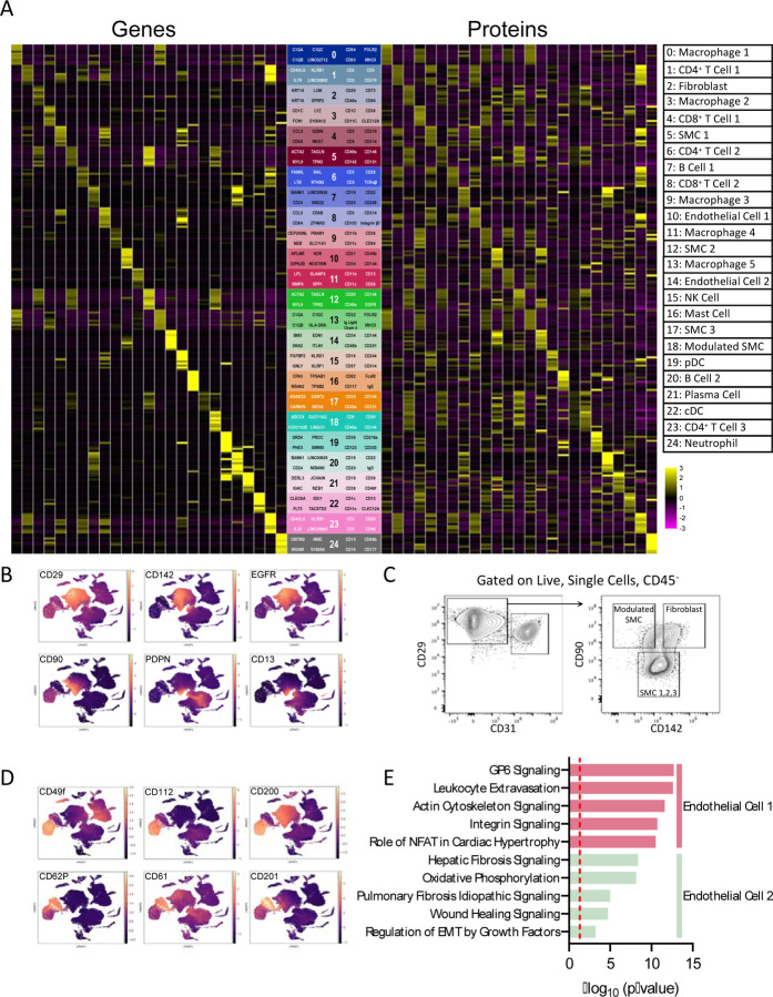
To characterize the landscape of human carotid atherosclerosis in greater detail, we combined cellular indexing of transcriptomes and epitopes by sequencing (CITE-seq) and single-cell RNA sequencing (scRNA-seq) to classify all cell types within lesions (n=21; 13 symptomatic) to achieve a comprehensive multimodal understanding of the cellular identities of atherosclerosis and their association with clinical pathophysiology.
We identified 25 distinct cell populations each having a unique multi-omic signature, including macrophages, T cells, NK cells, mast cells, B cells, plasma cells, neutrophils, dendritic cells, endothelial cells, fibroblasts, and smooth muscle cells (SMCs). Within the macrophage populations, we identified 2 proinflammatory subsets that were enriched in IL1B or C1Q expression, 2 distinct TREM2 positive foam cell subsets, one of which also expressed inflammatory genes, as well as subpopulations displaying a proliferative gene expression signature and one expressing SMC-specific genes and upregulation of fibrotic pathways. An in-depth characterization uncovered several subsets of SMCs and fibroblasts, including a SMC-derived foam cell. We localized this foamy SMC to the deep intima of coronary atherosclerotic lesions. Using CITE-seq data, we also developed the first flow cytometry panel, using cell surface proteins CD29, CD142, and CD90, to isolate SMC-derived cells from lesions. Last, we found that the proportion of efferocytotic macrophages, classically activated endothelial cells, contractile and modulated SMC-derived cell types were reduced, and inflammatory SMCs were enriched in plaques of clinically symptomatic vs. asymptomatic patients.
Our multimodal atlas of cell populations within atherosclerosis provides novel insights into the diversity, phenotype, location, isolation, and clinical relevance of the unique cellular composition of human carotid atherosclerosis. This facilitates both the mapping of cardiovascular disease susceptibility loci to specific cell types as well as the identification of novel molecular and cellular therapeutic targets for treatment of the disease.
动脉粥样硬化斑块是由细胞异质性混合物组成的复杂组织。然而,我们对这些病变内细胞的全面转录和表型格局了解有限。
为了更详细地表征人类颈动脉粥样硬化的格局,我们结合了通过测序进行转录组和表位的细胞索引(CITE-seq)和单细胞RNA测序(scRNA-seq),对病变内的所有细胞类型(n = 21;13例有症状)进行分类,以全面多模态地了解动脉粥样硬化的细胞身份及其与临床病理生理学的关联。
我们鉴定出25个不同的细胞群,每个细胞群都有独特的多组学特征,包括巨噬细胞、T细胞、自然杀伤细胞、肥大细胞、B细胞、浆细胞、中性粒细胞、树突状细胞、内皮细胞、成纤维细胞和平滑肌细胞(SMC)。在巨噬细胞群中,我们鉴定出2个促炎亚群,其IL1B或C1Q表达富集,2个不同的TREM2阳性泡沫细胞亚群,其中一个也表达炎症基因,以及显示增殖基因表达特征的亚群和一个表达SMC特异性基因并上调纤维化途径的亚群。深入表征发现了SMC和成纤维细胞的几个亚群,包括一个SMC衍生的泡沫细胞。我们将这种泡沫状SMC定位到冠状动脉粥样硬化病变的深层内膜。利用CITE-seq数据,我们还开发了第一个流式细胞术面板,使用细胞表面蛋白CD29、CD142和CD90,从病变中分离SMC衍生的细胞。最后,我们发现,与无症状患者相比,有临床症状患者的斑块中,具有吞噬功能的巨噬细胞、经典激活的内皮细胞、收缩性和调节性SMC衍生细胞类型的比例降低,而炎症性SMC富集。
我们的动脉粥样硬化内细胞群多模态图谱为人类颈动脉粥样硬化独特细胞组成的多样性、表型、位置、分离及临床相关性提供了新见解。这有助于将心血管疾病易感基因座映射到特定细胞类型,以及识别治疗该疾病的新型分子和细胞治疗靶点。