Yang Jae, Vrbovská Veronika, Franke Tilman, Sibert Bryan, Larson Matthew, Hall Alex, Rigort Alex, Mitchels John, Wright Elizabeth R
Department of Biochemistry, University of Wisconsin, Madison, WI USA.
Midwest Center for Cryo-Electron Tomography, Department of Biochemistry, University of Wisconsin, Madison, WI USA.
bioRxiv. 2023 Jul 12:2023.07.11.548578. doi: 10.1101/2023.07.11.548578.
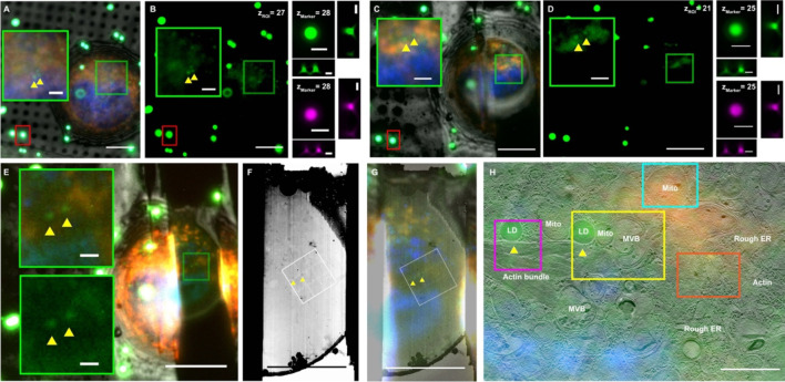
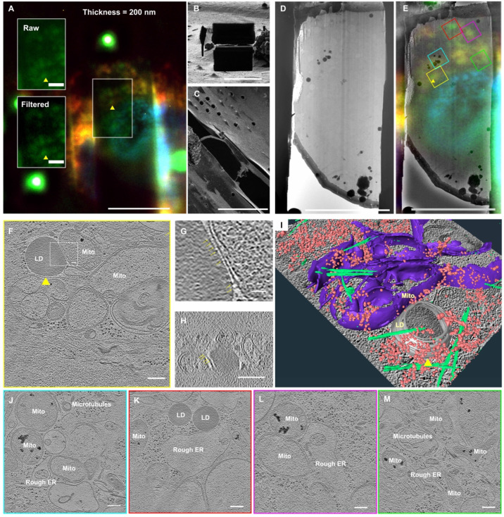
Correlative cryo-FLM-FIB milling is a powerful sample preparation technique for cryo-ET. However, correlative workflows that incorporate precise targeting remain challenging. Here, we demonstrate the development and use of an integrated Fluorescence Light Microscope (iFLM) module within a cryo-FIB-SEM to enable a coordinate-based two-point 3D correlative workflow. The iFLM guided targeting of regions of interest coupled with an automated milling process of the cryo-FIB-SEM instrument allows for the efficient preparation of 9-12 ∼200 nm thick lamellae within 24 hours. Using regular and montage-cryo-ET data collection schemes, we acquired data from FIB-milled lamellae of HeLa cells to examine cellular ultrastructure. Overall, this workflow facilitates on-the-fly targeting and automated FIB-milling of cryo-preserved cells, bacteria, and possibly high pressure frozen tissue, to produce lamellae for downstream cryo-ET data collection.
相关低温荧光光镜-聚焦离子束铣削是一种用于低温电子断层扫描的强大样品制备技术。然而,包含精确靶向的相关工作流程仍然具有挑战性。在这里,我们展示了在低温聚焦离子束扫描电子显微镜中开发和使用集成荧光显微镜(iFLM)模块,以实现基于坐标的两点三维相关工作流程。iFLM引导对感兴趣区域的靶向,再结合低温聚焦离子束扫描电子显微镜仪器的自动铣削过程,能够在24小时内高效制备出9至12个约200纳米厚的薄片。使用常规和拼接低温电子断层扫描数据采集方案,我们从HeLa细胞的聚焦离子束铣削薄片中获取数据,以检查细胞超微结构。总体而言,该工作流程有助于对冷冻保存的细胞、细菌以及可能的高压冷冻组织进行实时靶向和自动聚焦离子束铣削,以制备用于下游低温电子断层扫描数据采集的薄片。