MRC Laboratory of Molecular Biology, Cambridge CB2 0QH, UK.
MRC Laboratory of Molecular Biology, Cambridge CB2 0QH, UK.
Mol Cell. 2023 Aug 17;83(16):2911-2924.e16. doi: 10.1016/j.molcel.2023.06.035. Epub 2023 Jul 27.
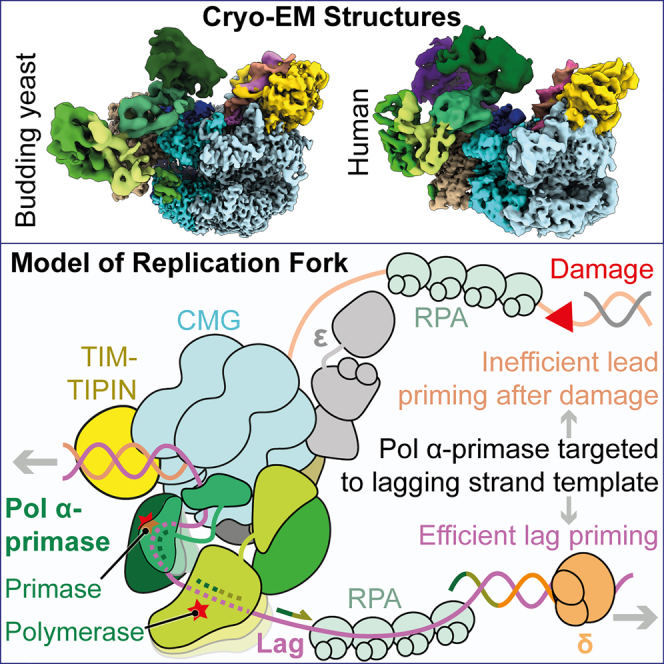
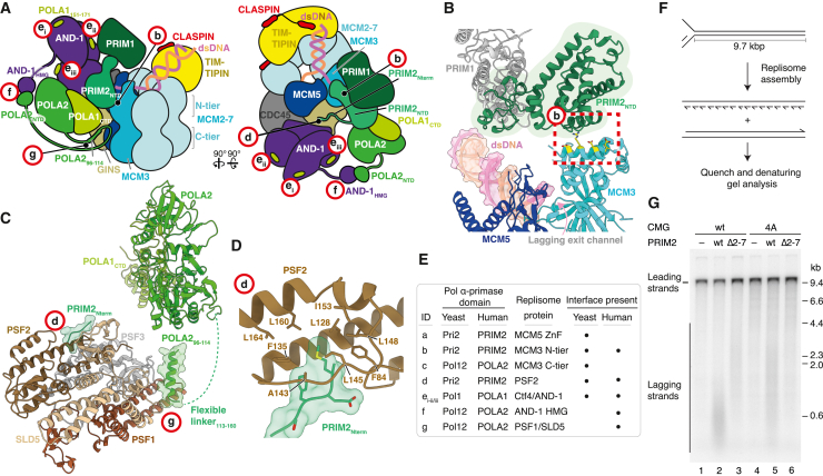
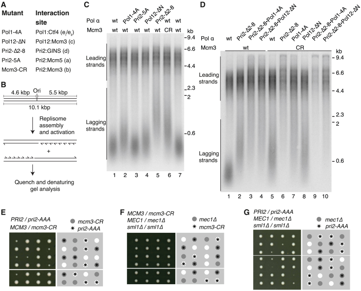
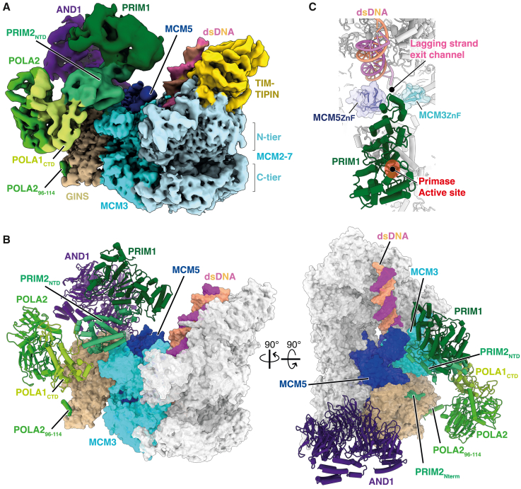
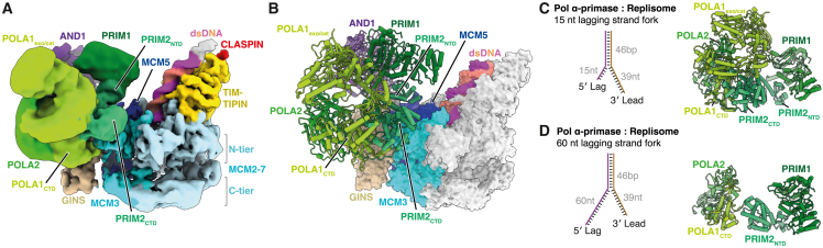
During eukaryotic DNA replication, Pol α-primase generates primers at replication origins to start leading-strand synthesis and every few hundred nucleotides during discontinuous lagging-strand replication. How Pol α-primase is targeted to replication forks to prime DNA synthesis is not fully understood. Here, by determining cryoelectron microscopy (cryo-EM) structures of budding yeast and human replisomes containing Pol α-primase, we reveal a conserved mechanism for the coordination of priming by the replisome. Pol α-primase binds directly to the leading edge of the CMG (CDC45-MCM-GINS) replicative helicase via a complex interaction network. The non-catalytic PRIM2/Pri2 subunit forms two interfaces with CMG that are critical for in vitro DNA replication and yeast cell growth. These interactions position the primase catalytic subunit PRIM1/Pri1 directly above the exit channel for lagging-strand template single-stranded DNA (ssDNA), revealing why priming occurs efficiently only on the lagging-strand template and elucidating a mechanism for Pol α-primase to overcome competition from RPA to initiate primer synthesis.
在真核生物 DNA 复制过程中,聚合酶 α-引发酶在复制起始点生成引物,以启动领头链合成,并在不连续的滞后链复制过程中每隔几百个核苷酸进行一次。然而,聚合酶 α-引发酶如何靶向复制叉以启动 DNA 合成,目前还不完全清楚。在这里,通过确定含有聚合酶 α-引发酶的芽殖酵母和人类复制体的冷冻电镜(cryo-EM)结构,我们揭示了复制体协调引发的保守机制。聚合酶 α-引发酶通过复杂的相互作用网络直接与 CMG(CDC45-MCM-GINS)复制解旋酶的前缘结合。非催化性 PRIM2/Pri2 亚基与 CMG 形成两个界面,对于体外 DNA 复制和酵母细胞生长至关重要。这些相互作用将引发酶的催化亚基 PRIM1/Pri1 直接定位在滞后链模板单链 DNA(ssDNA)的出口通道上方,解释了为什么引发只能在滞后链模板上有效进行,并阐明了聚合酶 α-引发酶克服 RPA 竞争以启动引物合成的机制。