Prasad Umakant, Suman Sanjay K, Kumari Manisha, Waghmare Vaibhav
Radiodiagnosis, Indira Gandhi Institute of Medical Sciences (IGIMS), Patna, IND.
Radiology, Indira Gandhi Institute of Medical Sciences (IGIMS), Patna, IND.
Cureus. 2023 Jun 25;15(6):e40932. doi: 10.7759/cureus.40932. eCollection 2023 Jun.
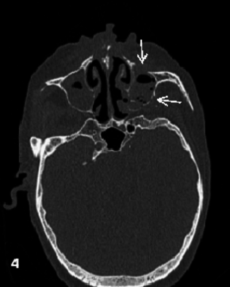
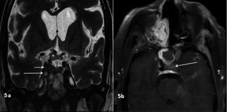
Aim We aim to study the spectrum of imaging findings in patients with rhino-oculo-cerebral mucormycosis (ROCM). Materials and methods This retrospective descriptive study was performed in histopathologically confirmed cases of rhino-oculo-cerebral mucormycosis in a tertiary care center in Bihar, India. The case records of patients with radiological, cultural, and histological evidence of acute invasive ROCM were retrospectively evaluated for relevant radiological and clinical data between May 2021 and June 2022. Results The radiological evaluation included computed tomography (CT) and magnetic resonance imaging (MRI) scans done on 52 patients. The patient's average age was 48 years. The ethmoid sinus was involved in 51 (98%) cases and the maxillary sinus in 50 (96%) cases. Bilateral sinus involvement (45, 86%) was the most common, followed by pansinus involvement (27, 52%). The orbit was involved in 39 (75%) cases, the face in 25 (47%) cases, and retroantral fat stranding in 24 (46%) cases. Mucosal thickening (91%) was the most common pattern of involvement, followed by complete opacification (77%). Osseous involvement was seen in 17 of 44 patients who had CT scans, and the majority of patients had extrasinus extension with intact bone. MRI revealed variable T2SI, with T2 hyperintensity being the most common pattern. Heterogeneous enhancement in post-contrast imaging was the most common. Conclusion ROCM is a life-threatening invasive fungal infection, especially in an immunocompromised state. ROCM is characterized by a variety of imaging abnormalities on CT and MRI, although nonspecific. Imaging aids in suspicion or early diagnosis in appropriate clinical contexts, particularly in an immunocompromised state, and in determining the degree of involvement and complications. Early detection of ROCM and its complications enables proper treatment, which can lower the cost of care, morbidity, and mortality.
目的 我们旨在研究鼻眼脑型毛霉菌病(ROCM)患者的影像学表现谱。
材料与方法 本回顾性描述性研究在印度比哈尔邦一家三级医疗中心对组织病理学确诊的鼻眼脑型毛霉菌病病例进行。对2021年5月至2022年6月期间有急性侵袭性ROCM放射学、培养及组织学证据的患者病历进行回顾性评估,以获取相关放射学和临床数据。
结果 放射学评估包括对52例患者进行的计算机断层扫描(CT)和磁共振成像(MRI)扫描。患者平均年龄为48岁。筛窦受累51例(98%),上颌窦受累50例(96%)。双侧鼻窦受累(45例,86%)最为常见,其次是全鼻窦受累(27例,52%)。眼眶受累39例(75%),面部受累25例(47%),窦后脂肪条索征24例(46%)。黏膜增厚(91%)是最常见的受累模式,其次是完全闭塞(77%)。44例行CT扫描的患者中有17例出现骨质受累,大多数患者鼻窦外扩展且骨质完整。MRI显示T2信号强度各异,T2高信号是最常见的模式。增强后成像中不均匀强化最为常见。
结论 ROCM是一种危及生命的侵袭性真菌感染,尤其是在免疫功能低下状态下。ROCM的特征是CT和MRI上有多种影像学异常,尽管不具有特异性。影像学有助于在适当的临床背景下,特别是在免疫功能低下状态下进行怀疑或早期诊断,并有助于确定受累程度和并发症。早期发现ROCM及其并发症能够进行恰当治疗,从而降低医疗成本、发病率和死亡率。