Department of Radiology, Seth GSMC & KEMH, Parel, Mumbai 400012, India.
Department of Radiology, Seth GSMC & KEMH, Parel, Mumbai 400012, India.
Clin Imaging. 2022 Feb;82:172-178. doi: 10.1016/j.clinimag.2021.10.014. Epub 2021 Nov 6.
The study aims to depict the radiological features of Cov-ROCM, depict the common routes of spread to orbits and intracranial compartment and look for an association of the risk factors with radiological severity of the disease.
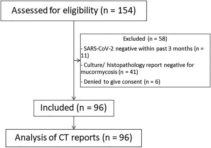
96 patients who had COVID-19 infection in the past 3 months and were diagnosed with ROCM underwent CECT PNS examinations which were assessed by two experienced radiologists. They were divided into three groups based on the intraorbital and intracranial involvement and were correlated with various risk factors.
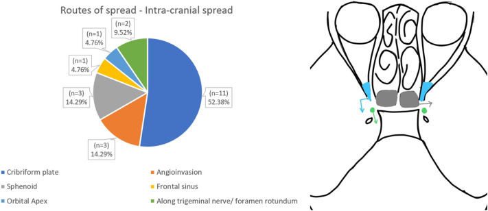
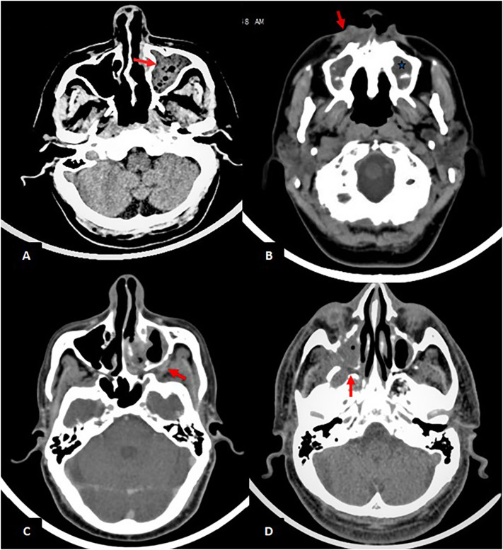
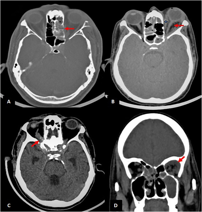
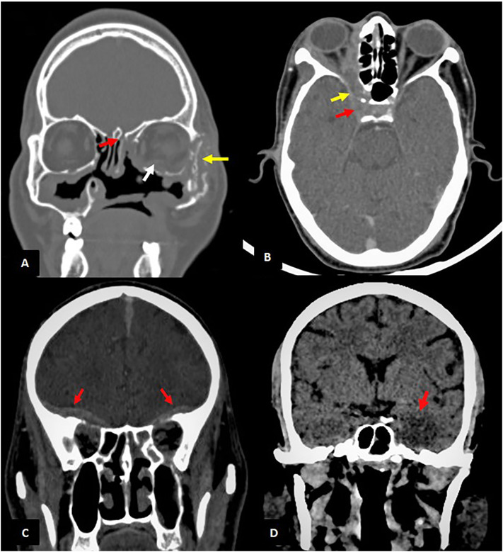
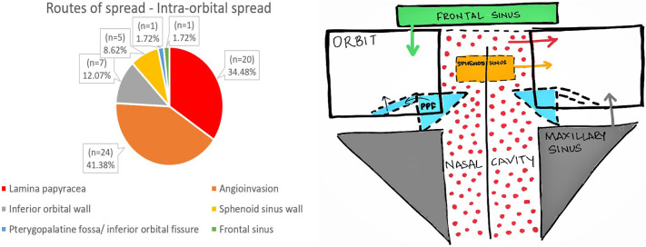
The incidence of bony erosions which was the commonest finding (75%) was double in Cov-ROCM than the ROCM cases of pre COVID era (33-40%). The most common route of spread to orbit was through angioinvasion(52%) with intact orbital walls; and intracranial extension was via erosion of the cribriform plate(52%). Sphenoid sinus involvement is strongly associated with intracranial and intraorbital involvement.(p-value = .0004). History of longer ICU stays and being on mechanical ventilation as a part of COVID management is associated with aggressive disease pattern(p-value = .002). Similarly, poor glycaemic control signified by raised HbA1c levels showed statistically significant correlation with severe Cov-ROCM(intraorbital/intracranial extension) (p-value = .040).
Amidst the COVID pandemic, it is pertinent to look at bony erosions in case of any sinusitis, especially bony maxillary walls and the turbinates. The intraorbital compartment must be viewed thoroughly even in the absence of bony erosions due to the angioinvasive nature of these fungi. Aggressive follow-up for patients with ICU stays for COVID and for glycaemic control would help reduce the morbidity.
本研究旨在描述 Cov-ROCM 的放射学特征,描述常见的眼眶和颅内蔓延途径,并寻找与疾病放射学严重程度相关的危险因素。
对过去 3 个月内患有 COVID-19 感染并诊断为 ROCM 的 96 例患者进行 CECT PNS 检查,由两位有经验的放射科医生进行评估。根据眼眶和颅内受累情况将其分为三组,并与各种危险因素相关联。
骨侵蚀(最常见的表现,发生率为 75%)在 Cov-ROCM 中比 COVID 前时代(33-40%)的 ROCM 病例更为常见。最常见的眼眶蔓延途径是通过血管侵犯(52%),眼眶壁完整;颅内延伸是通过筛板侵蚀(52%)。蝶窦受累与颅内和眼眶受累密切相关(p 值=0.0004)。在 COVID 管理中,ICU 停留时间较长和机械通气史与侵袭性疾病模式相关(p 值=0.002)。同样,糖化血红蛋白水平升高表明血糖控制不佳,与严重的 Cov-ROCM(眼眶/颅内延伸)有统计学显著相关性(p 值=0.040)。
在 COVID 大流行期间,对于任何鼻窦炎,特别是上颌骨壁和鼻甲的骨侵蚀,都应进行影像学检查。由于这些真菌具有血管侵犯性,即使没有骨侵蚀,也必须仔细观察眼眶间隙。对于因 COVID 入住 ICU 的患者和控制血糖的患者进行积极的随访,有助于降低发病率。