Department of Molecular and Cell Biology, University of California, Berkeley, CA 94720.
Helen Wills Neuroscience Institute, University of California, Berkeley, CA 94720.
Proc Natl Acad Sci U S A. 2023 Aug 8;120(32):e2221122120. doi: 10.1073/pnas.2221122120. Epub 2023 Jul 31.
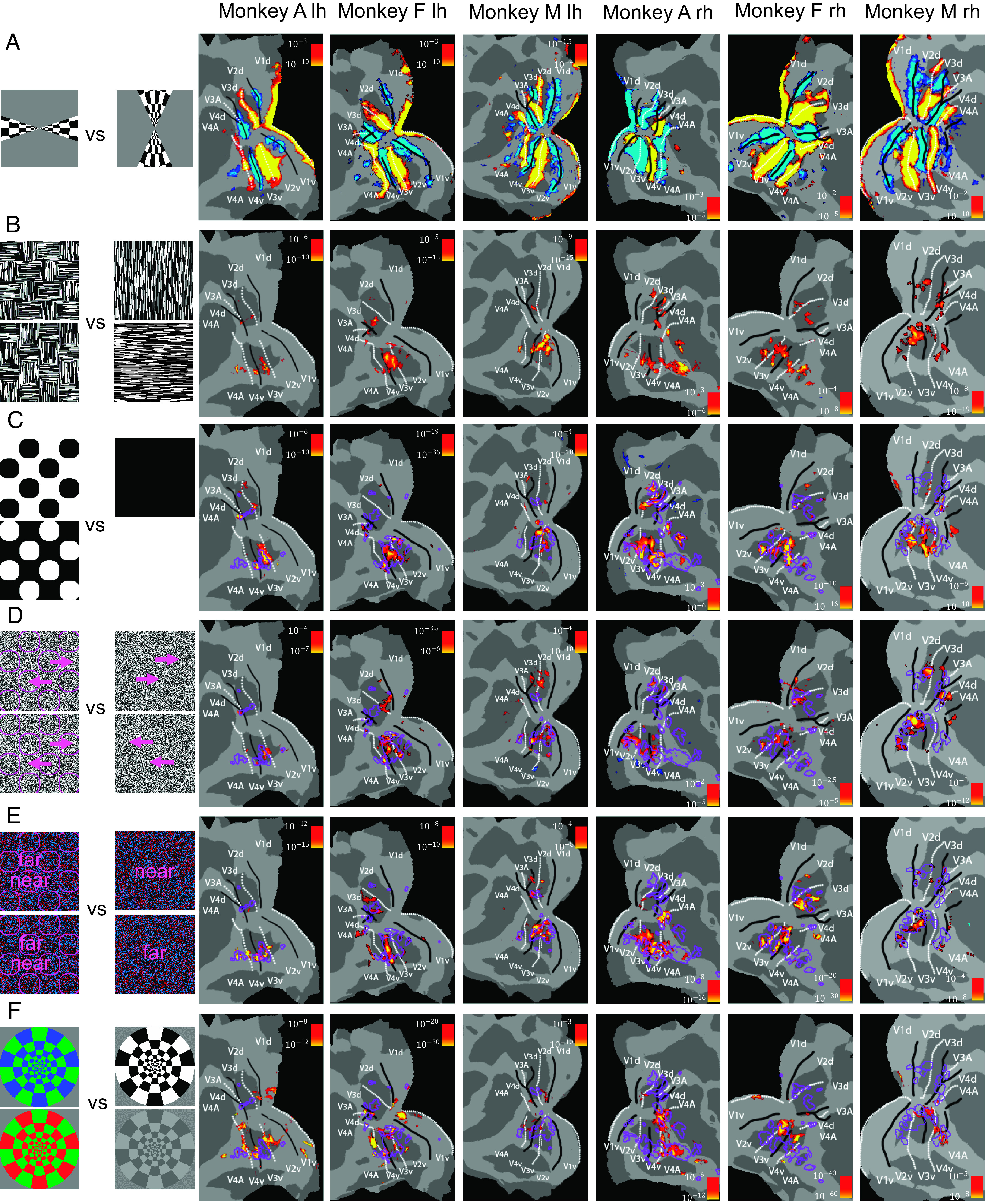
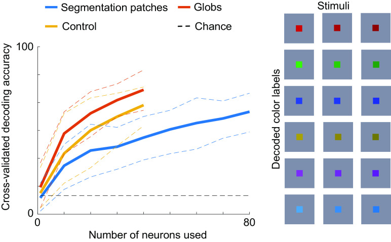
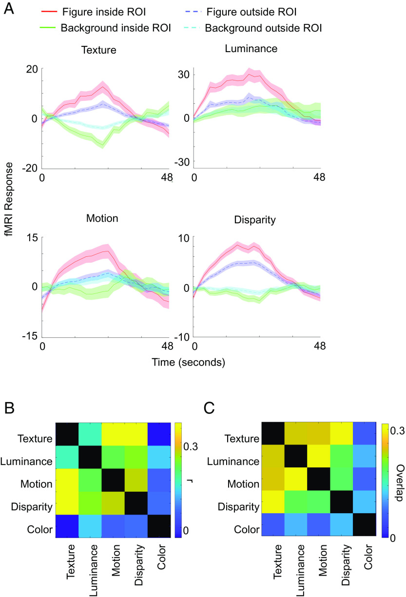
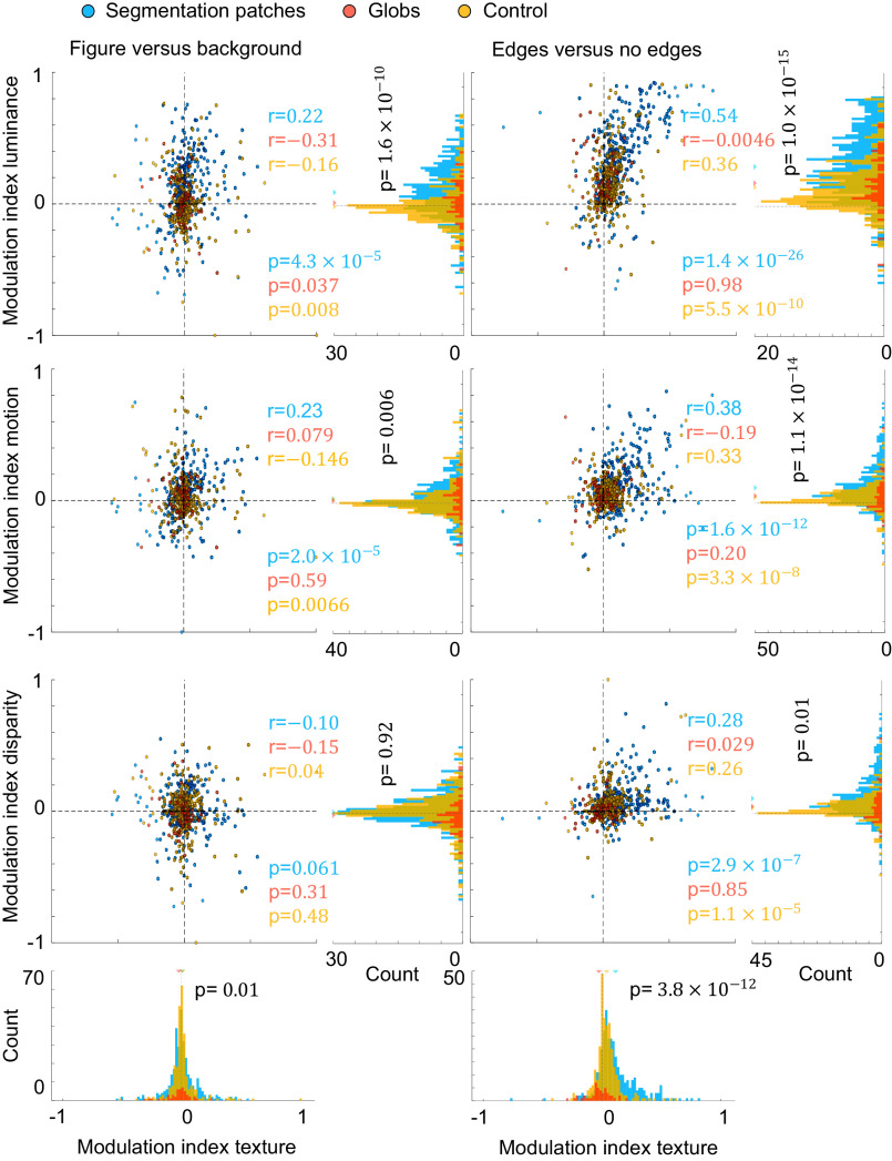
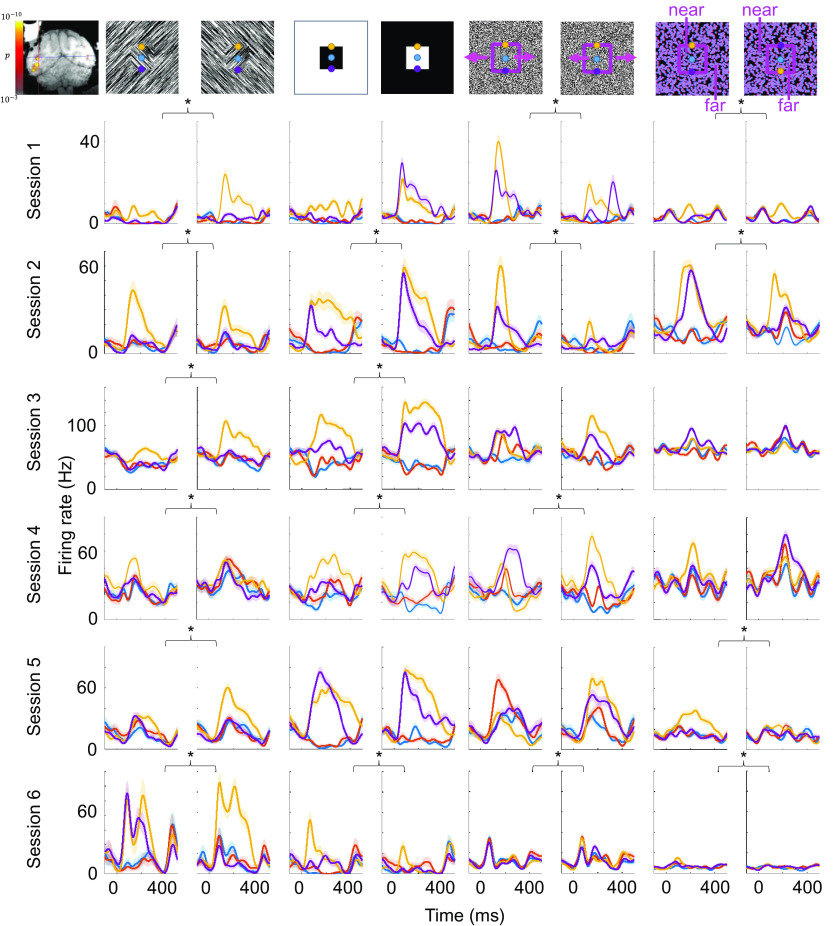
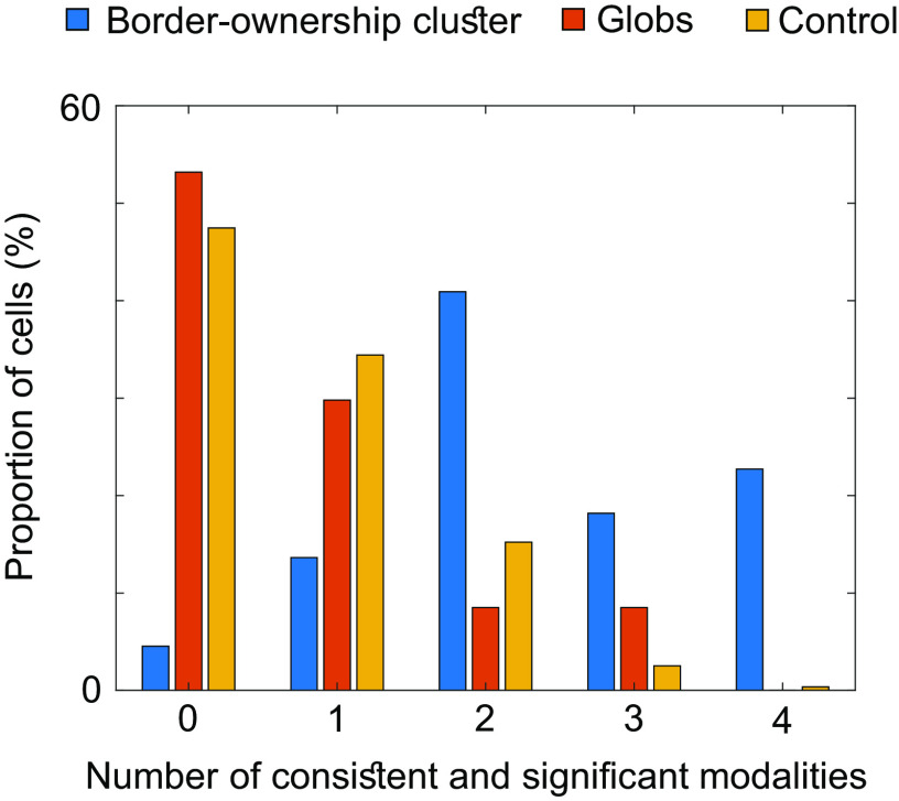
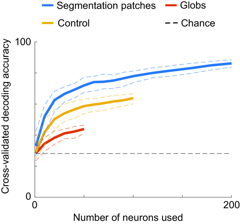
Segmentation, the computation of object boundaries, is one of the most important steps in intermediate visual processing. Previous studies have reported cells across visual cortex that are modulated by segmentation features, but the functional role of these cells remains unclear. First, it is unclear whether these cells encode segmentation consistently since most studies used only a limited variety of stimulus types. Second, it is unclear whether these cells are organized into specialized modules or instead randomly scattered across the visual cortex: the former would lend credence to a functional role for putative segmentation cells. Here, we used fMRI-guided electrophysiology to systematically characterize the consistency and spatial organization of segmentation-encoding cells across the visual cortex. Using fMRI, we identified a set of patches in V2, V3, V3A, V4, and V4A that were more active for stimuli containing figures compared to ground, regardless of whether figures were defined by texture, motion, luminance, or disparity. We targeted these patches for single-unit recordings and found that cells inside segmentation patches were tuned to both figure-ground and borders more consistently across types of stimuli than cells in the visual cortex outside the patches. Remarkably, we found clusters of cells inside segmentation patches that showed the same border-ownership preference across all stimulus types. Finally, using a population decoding approach, we found that segmentation could be decoded with higher accuracy from segmentation patches than from either color-selective or control regions. Overall, our results suggest that segmentation signals are preferentially encoded in spatially discrete patches.
分割,即计算物体边界,是中间视觉处理中最重要的步骤之一。先前的研究报告了跨视觉皮层的细胞受到分割特征的调节,但这些细胞的功能作用仍不清楚。首先,由于大多数研究仅使用有限种类的刺激类型,因此不清楚这些细胞是否一致地编码分割。其次,不清楚这些细胞是否组织成专门的模块,或者随机分散在视皮层中:前者将为假定的分割细胞的功能作用提供依据。在这里,我们使用 fMRI 引导的电生理学方法系统地描述了整个视皮层中分割编码细胞的一致性和空间组织。使用 fMRI,我们在 V2、V3、V3A、V4 和 V4A 中确定了一组斑块,这些斑块对于包含图形的刺激比地面更活跃,无论图形是由纹理、运动、亮度还是视差定义的。我们针对这些斑块进行了单细胞记录,发现与斑块外的视觉皮层中的细胞相比,斑块内的细胞在所有刺激类型中对图形-背景和边界的调谐更一致。值得注意的是,我们发现斑块内的细胞簇在所有刺激类型中都表现出相同的边界所有权偏好。最后,使用群体解码方法,我们发现从分割斑块中可以比从颜色选择或控制区域中更准确地解码分割。总的来说,我们的结果表明,分割信号优先在空间离散的斑块中进行编码。