Department of Oral and Maxillofacial Surgery, University Hospital Carl Gustav Carus, 01304, Dresden, Germany.
Department of Diagnostic and Interventional Neuroradiology, University Hospital Carl Gustav Carus, Dresden, Germany.
Clin Oral Investig. 2023 Sep;27(9):5637-5647. doi: 10.1007/s00784-023-05185-x. Epub 2023 Aug 3.
Symmetry is essential for computer-aided surgical (CAS) procedures in oral and maxillofacial surgery (OMFS). A critical step for successful CAS is mirroring the unaffected side to create a template for the virtual reconstruction of the injured anatomical structure. The aim was to identify specific anatomical landmarks of the midfacial skeleton, to evaluate the symmetry in a group of the real-world Central European population, and to use these landmarks to assess midfacial symmetry in CT scans.
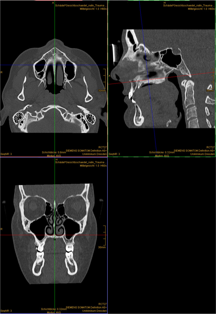
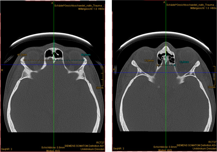
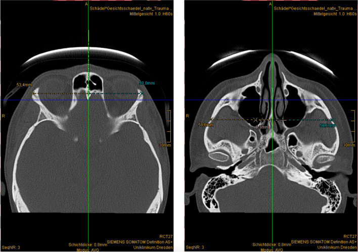
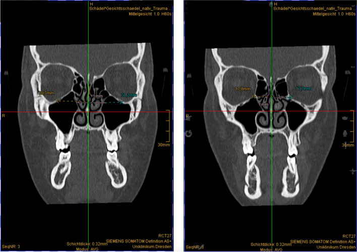
The retrospective cross-sectional study defined landmarks of the midface's bony contour using viscerocranial CT data. The distances of the skeletal landmarks (e.g., the frontozygomatic suture and temporozygomatic suture) of the left and right sides from the midline were measured and statistically compared. Midfacial symmetry for reference points was defined as a difference within 0 mm and their mean difference plus one standard deviation.
We examined a total of 101 CT scans. 75% of our population shows symmetrical proportions of the midface. The means of the differences for the left and right sides ranged from 0.8 to 1.3 mm, averaging 1.1 ± 0.2 mm for all skeletal landmarks. The standard deviations ranged from 0.6 to 1.4 mm, with a computed mean of 0.9 ± 0.3 mm.
We established a methodology to assess the symmetry of the bony midface. If the determined differences were equal to or lower than 2.5 mm in the mentioned midfacial skeletal landmarks, then the symmetry of the bony midface was considered present, and symmetry-based methods for CAS procedures are applicable.
Many CAS procedures require facial symmetry. We provide an easy-to-apply method to probe for symmetry of the midface. The method may be used for population-based research, to check for proper reduction of fractures after reposition or to screen for symmetry prior to CAS planning.
对称性对于口腔颌面外科(OMFS)中的计算机辅助手术(CAS)至关重要。成功进行 CAS 的关键步骤是镜像未受影响的一侧,为受伤解剖结构的虚拟重建创建模板。本研究旨在确定面中部骨骼的特定解剖标志,评估一组中欧真实人群的对称性,并使用这些标志评估 CT 扫描中的面中部对称性。
本回顾性横断面研究使用内脏颅 CT 数据对面中部骨骼轮廓的标志进行定义。测量左右侧骨骼标志(例如额颧缝和颞颧缝)与中线的距离,并进行统计学比较。参考点的面中部对称性定义为差异在 0mm 以内及其平均值加一个标准差。
我们共检查了 101 份 CT 扫描。我们人群的 75%显示出面中部比例对称。左右两侧差异的平均值范围为 0.8 至 1.3mm,所有骨骼标志的平均值为 1.1 ± 0.2mm。标准差范围为 0.6 至 1.4mm,计算平均值为 0.9 ± 0.3mm。
我们建立了一种评估骨性面中部对称性的方法。如果在提到的面中部骨骼标志中确定的差异等于或低于 2.5mm,则认为骨性面中部存在对称性,并且适用于基于对称性的 CAS 程序方法。
许多 CAS 程序都需要面部对称性。我们提供了一种易于应用的方法来探测面中部的对称性。该方法可用于基于人群的研究,以检查骨折复位后的适当复位情况,或在 CAS 计划之前进行对称性筛查。