van den Bulk Jitske, Verdegaal Els M E, van der Ploeg Manon, Visser Marten, Nunes Joana B, de Ru Arnoud H, Tjokrodirijo Rayman T N, Ijsselsteijn Marieke E, Janssen Natasja I, van der Breggen Ruud, de Bruin Linda, de Kok Pita, Janssen George M C, Ruano Dina, Kapiteijn Ellen H W, van Veelen Peter A, de Miranda Noel F C C, van der Burg Sjoerd H
Department of Pathology, Leiden University Medical Center, Leiden, the Netherlands.
Department of Medical Oncology, Oncode Institute, Leiden University Medical Center, Leiden, the Netherlands.
Clin Cancer Res. 2023 Oct 13;29(20):4278-4288. doi: 10.1158/1078-0432.CCR-23-1106.
The availability of (neo)antigens and the infiltration of tumors by (neo)antigen-specific T cells are crucial factors in cancer immunotherapy. In this study, we aimed to investigate the targetability of (neo)antigens in advanced progessive melanoma and explore the potential for continued T-cell-based immunotherapy.
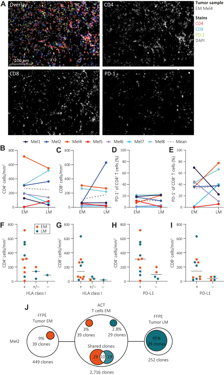
We examined a cohort of eight patients with melanoma who had sequential metastases resected at early and later time points. Antigen-presenting capacity was assessed using IHC and flow cytometry. T-cell infiltration was quantified through multiplex immunofluorescence. Whole-exome and RNA sequencing were conducted to identify neoantigens and assess the expression of neoantigens and tumor-associated antigens. Mass spectrometry was used to evaluate antigen presentation. Tumor recognition by autologous T cells was assessed by coculture assays with cell lines derived from the metastatic lesions.
We observed similar T-cell infiltration in paired early and later metastatic (LM) lesions. Although elements of the antigen-presenting machinery were affected in some LM lesions, both the early and later metastasis-derived cell lines were recognized by autologous T cells. At the genomic level, the (neo)antigen landscape was dynamic, but the (neo)antigen load was stable between paired lesions.
Our findings indicate that subsequently isolated tumors from patients with late-stage melanoma retain sufficient antigen-presenting capacity, T-cell infiltration, and a stable (neo)antigen load, allowing recognition of tumor cells by T cells. This indicates a continuous availability of T-cell targets in metastases occurring at different time points and supports further exploration of (neo)antigen-specific T-cell-based therapeutic approaches for advanced melanoma.
(新)抗原的可用性以及(新)抗原特异性T细胞对肿瘤的浸润是癌症免疫治疗的关键因素。在本研究中,我们旨在调查晚期进展性黑色素瘤中(新)抗原的靶向性,并探索基于T细胞的免疫治疗的潜力。
我们检查了一组八名黑色素瘤患者,他们在早期和晚期时间点先后切除了转移性肿瘤。使用免疫组化和流式细胞术评估抗原呈递能力。通过多重免疫荧光对T细胞浸润进行定量。进行全外显子组和RNA测序以鉴定新抗原,并评估新抗原和肿瘤相关抗原的表达。使用质谱法评估抗原呈递。通过与源自转移性病变的细胞系共培养试验评估自体T细胞对肿瘤的识别。
我们在配对的早期和晚期转移性(LM)病变中观察到相似的T细胞浸润。尽管在一些LM病变中抗原呈递机制的成分受到影响,但早期和晚期转移来源的细胞系均被自体T细胞识别。在基因组水平上,(新)抗原格局是动态的,但配对病变之间的(新)抗原负荷是稳定的。
我们的研究结果表明,随后从晚期黑色素瘤患者中分离出的肿瘤保留了足够的抗原呈递能力、T细胞浸润和稳定的(新)抗原负荷,使得T细胞能够识别肿瘤细胞。这表明在不同时间点发生的转移灶中持续存在T细胞靶点,并支持进一步探索针对晚期黑色素瘤的基于(新)抗原特异性T细胞的治疗方法。