• 文献检索
  • 文档翻译
  • 深度研究
  • 学术资讯
  • Suppr Zotero 插件Zotero 插件
  • 邀请有礼
  • 套餐&价格
  • 历史记录
应用&插件
Suppr Zotero 插件Zotero 插件浏览器插件Mac 客户端Windows 客户端微信小程序
定价
高级版会员购买积分包购买API积分包
服务
文献检索文档翻译深度研究API 文档MCP 服务
关于我们
关于 Suppr公司介绍联系我们用户协议隐私条款
关注我们

Suppr 超能文献

核心技术专利:CN118964589B侵权必究
粤ICP备2023148730 号-1Suppr @ 2026

文献检索

告别复杂PubMed语法,用中文像聊天一样搜索,搜遍4000万医学文献。AI智能推荐,让科研检索更轻松。

立即免费搜索

文件翻译

保留排版,准确专业,支持PDF/Word/PPT等文件格式,支持 12+语言互译。

免费翻译文档

深度研究

AI帮你快速写综述,25分钟生成高质量综述,智能提取关键信息,辅助科研写作。

立即免费体验

Spitz痣与黑色素瘤:皮肤镜及反射式共聚焦显微镜评估

Spitz nevus and melanoma: evaluation with dermoscopy and reflectance confocal microscopy.

作者信息

Pogorzelska-Antkowiak Anna, Slowinska Monika, Farinazzo Eleonora, Conforti Claudio, Agozzino Marina

机构信息

EsteDerm Private Dermatology Clinic, Tychy, Poland.

Military Hospital, Warsaw, Poland.

出版信息

Postepy Dermatol Alergol. 2023 Jun;40(3):427-431. doi: 10.5114/ada.2023.125968. Epub 2023 Mar 21.

DOI:10.5114/ada.2023.125968
PMID:37545824
原文链接:https://pmc.ncbi.nlm.nih.gov/articles/PMC10399674/
Abstract

INTRODUCTION

Spitz nevi (SN) include a wide range of benign melanocytic nevi, which are controversial due to their morphologic resemblance to melanoma (MM).

AIM

To describe dermoscopic and reflectance confocal microscopic (RCM) features of SN compared to MM and assess the RCM utility in the differential diagnosis.

MATERIAL AND METHODS

We performed a multicentre retrospective analysis of MM and SN evaluated with dermoscopy and reflectance confocal microscopy. Three RCM mosaics were obtained for each lesion. Nine dermoscopic and twenty-one microscopic features were assessed for each lesion.

RESULTS

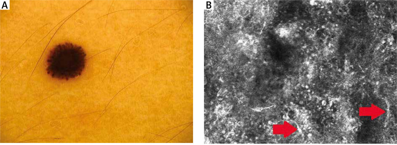
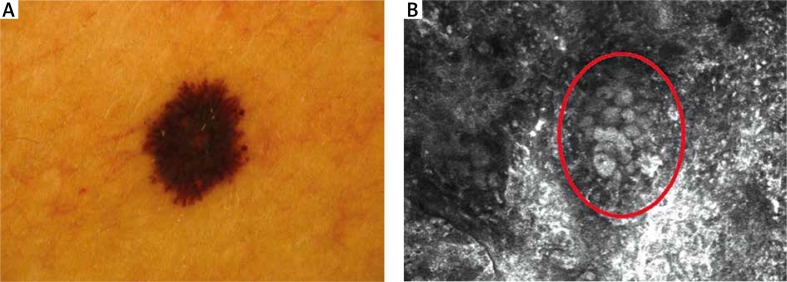
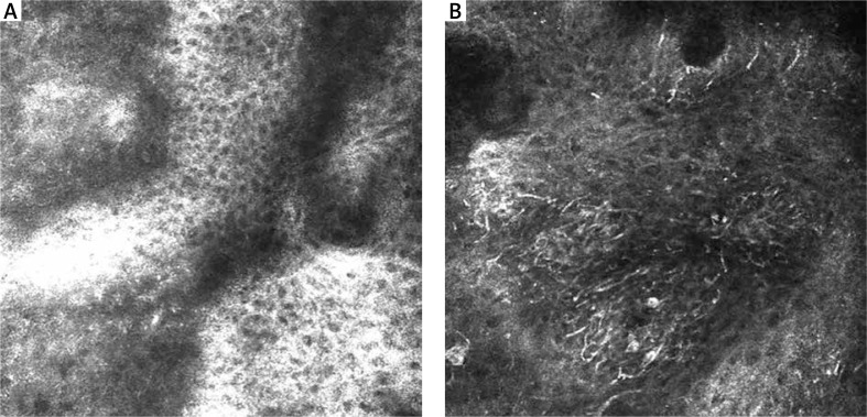
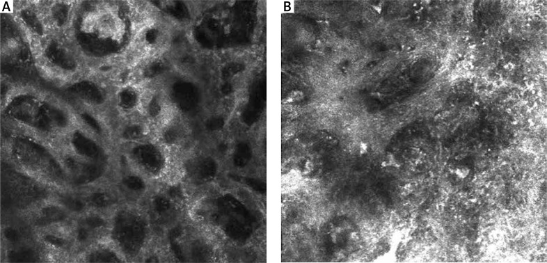
A total of 26 lesions (15 SN and 11 MM) were included. Dermoscopically, most SN showed a "starburst" pattern. Asymmetry was marked in 8 MM. There were 6 dermoscopic features significantly more prevalent in MM than in SN. RCM showed that an atypical honeycomb pattern, atypical infiltration, and disarray of the epidermis were significant for MM. SN mostly revealed a typical honeycomb pattern. At the DEJ, most of SN had a meshwork pattern; MM revealed an atypical meshwork pattern. Atypical cells and sheet-like structures were observed in most MM.

CONCLUSIONS

A combined approach using dermoscopy and RCM supports the differential diagnosis of SN and MM. Although our study showed some significant differences between SN and MM in RCM, further research on a larger group should be considered.

摘要

引言

斯皮茨痣(SN)包括多种良性黑素细胞痣,因其形态与黑色素瘤(MM)相似而存在争议。

目的

描述斯皮茨痣与黑色素瘤相比的皮肤镜和反射式共聚焦显微镜(RCM)特征,并评估RCM在鉴别诊断中的效用。

材料与方法

我们对采用皮肤镜和反射式共聚焦显微镜评估的黑色素瘤和斯皮茨痣进行了多中心回顾性分析。每个病变获取三张RCM拼接图。对每个病变评估九种皮肤镜特征和二十一种显微镜特征。

结果

共纳入26个病变(15个斯皮茨痣和11个黑色素瘤)。在皮肤镜检查中,大多数斯皮茨痣表现为“星爆”模式。8个黑色素瘤表现出明显的不对称性。有6种皮肤镜特征在黑色素瘤中比在斯皮茨痣中更常见。RCM显示,非典型蜂巢状模式、非典型浸润和表皮紊乱对黑色素瘤具有重要意义。斯皮茨痣大多表现为典型的蜂巢状模式。在真皮表皮交界处,大多数斯皮茨痣具有网状模式;黑色素瘤表现为非典型网状模式。大多数黑色素瘤中观察到非典型细胞和片状结构。

结论

皮肤镜和RCM联合应用有助于斯皮茨痣和黑色素瘤的鉴别诊断。尽管我们的研究显示斯皮茨痣和黑色素瘤在RCM方面存在一些显著差异,但仍应考虑对更大样本量进行进一步研究。

https://cdn.ncbi.nlm.nih.gov/pmc/blobs/502f/10399674/5cd8f09c2b46/PDIA-40-50363-g004.jpg
https://cdn.ncbi.nlm.nih.gov/pmc/blobs/502f/10399674/933371954644/PDIA-40-50363-g001.jpg
https://cdn.ncbi.nlm.nih.gov/pmc/blobs/502f/10399674/7885afaad68e/PDIA-40-50363-g002.jpg
https://cdn.ncbi.nlm.nih.gov/pmc/blobs/502f/10399674/7722c8a010c2/PDIA-40-50363-g003.jpg
https://cdn.ncbi.nlm.nih.gov/pmc/blobs/502f/10399674/5cd8f09c2b46/PDIA-40-50363-g004.jpg

相似文献

1
Spitz nevus and melanoma: evaluation with dermoscopy and reflectance confocal microscopy.Spitz痣与黑色素瘤:皮肤镜及反射式共聚焦显微镜评估
Postepy Dermatol Alergol. 2023 Jun;40(3):427-431. doi: 10.5114/ada.2023.125968. Epub 2023 Mar 21.
2
Role of In Vivo Reflectance Confocal Microscopy in the Analysis of Melanocytic Lesions.体内反射共聚焦显微镜在黑素细胞性病变分析中的作用
Acta Dermatovenerol Croat. 2018 Apr;26(1):64-67.
3
Correlation of dermoscopic structures of melanocytic lesions to reflectance confocal microscopy.黑素细胞性皮损的皮肤镜结构与反射式共聚焦显微镜检查的相关性
Arch Dermatol. 2007 Feb;143(2):176-85. doi: 10.1001/archderm.143.2.176.
4
Reflectance confocal microscopy analysis of equivocal melanocytic lesions with severe regression.对伴有严重消退的可疑黑素细胞性病变进行反射式共聚焦显微镜分析。
Skin Res Technol. 2018 Feb;24(1):9-15. doi: 10.1111/srt.12382. Epub 2017 May 21.
5
In vivo confocal microscopy: The role of comparative approach in patients with multiple atypical nevi.体内共聚焦显微镜:比较方法在多发性非典型痣患者中的作用。
Exp Dermatol. 2020 Oct;29(10):945-952. doi: 10.1111/exd.14162. Epub 2020 Sep 21.
6
Dermoscopic and clinical predictors of reflectance confocal microscopy patterns of typical nevi on the back and legs: A cross-sectional study.背部和腿部典型痣的反射共聚焦显微镜模式的皮肤镜和临床预测因子:一项横断面研究。
J Am Acad Dermatol. 2021 Nov;85(5):1240-1247. doi: 10.1016/j.jaad.2020.06.020. Epub 2020 Jun 10.
7
Dendritic cells in reflectance confocal microscopy are a clue for early melanoma diagnosis in extrafacial flat pigmented melanocytic lesions.在非面部扁平色素性黑素细胞病变中,反射共聚焦显微镜下的树突状细胞是早期诊断黑素瘤的线索。
Exp Dermatol. 2022 Jul;31(7):1048-1055. doi: 10.1111/exd.14553. Epub 2022 Mar 4.
8
Acral pigmented Spitz nevus in a child with transepidermal migration of melanocytes: Dermoscopic and reflectance confocal microscopic features.一名黑素细胞发生表皮迁移的儿童的肢端色素性斯皮茨痣:皮肤镜及反射式共聚焦显微镜特征
Pediatr Dermatol. 2018 Mar;35(2):e99-e102. doi: 10.1111/pde.13385. Epub 2018 Jan 4.
9
In vivo reflectance confocal microscopy of halo nevi.活体共聚焦反射显微镜下观察晕痣。
Skin Res Technol. 2021 Sep;27(5):841-845. doi: 10.1111/srt.13029. Epub 2021 Mar 10.
10
Dynamic dermoscopic and reflectance confocal microscopic changes of melanocytic lesions excised during follow up.随访切除的黑素细胞病变的动态皮肤镜和反射共聚焦显微镜变化。
J Am Acad Dermatol. 2022 May;86(5):1049-1057. doi: 10.1016/j.jaad.2021.03.081. Epub 2021 Apr 3.

引用本文的文献

1
Clinical, Dermoscopic and Reflectance Confocal Microscopy Characteristics Associated With the Presence of Negative Pigment Network Among Spitzoid Neoplasms.与Spitzoid肿瘤中无色素网络存在相关的临床、皮肤镜及反射式共聚焦显微镜特征
Exp Dermatol. 2025 Aug;34(8):e70154. doi: 10.1111/exd.70154.

本文引用的文献

1
Dermatoscopy.皮肤镜检查。
Clin Dermatol. 2021 Jul-Aug;39(4):635-642. doi: 10.1016/j.clindermatol.2021.03.009. Epub 2021 Mar 19.
2
Mimics of melanoma in reflectance confocal microscopy.反射共聚焦显微镜下的黑色素瘤模拟物。
Int J Dermatol. 2021 May;60(5):540-546. doi: 10.1111/ijd.15306. Epub 2020 Nov 9.
3
Reflectance confocal microscopy characteristics for melanocytic nevi.黑素细胞痣的反射式共聚焦显微镜特征
Chin Med J (Engl). 2019 Oct 20;132(20):2510-2511. doi: 10.1097/CM9.0000000000000440.
4
Reevaluation of established and new criteria in differential diagnosis of Spitz nevus and melanoma.Spitz 痣与黑色素瘤鉴别诊断中既定和新准则的再评估。
Arch Dermatol Res. 2018 May;310(4):329-342. doi: 10.1007/s00403-018-1818-8. Epub 2018 Feb 7.
5
Dermoscopic Patterns of Spitz Nevi.Spitz痣的皮肤镜表现
JAMA. 2018 Jan 9;319(2):194. doi: 10.1001/jama.2017.18991.
6
Spitz Tumors of the Skin.皮肤的斯皮茨肿瘤
Surg Pathol Clin. 2017 Jun;10(2):281-298. doi: 10.1016/j.path.2017.01.004. Epub 2017 Mar 31.
7
The diagnosis and management of the Spitz nevus in the pediatric population: a systematic review and meta-analysis protocol.儿童人群中Spitz痣的诊断与管理:一项系统评价和Meta分析方案
Syst Rev. 2017 Apr 13;6(1):81. doi: 10.1186/s13643-017-0477-8.
8
Update on dermoscopy of Spitz/Reed naevi and management guidelines by the International Dermoscopy Society.Spitz/Reed 痣的皮肤镜检查更新和国际皮肤镜学会的管理指南。
Br J Dermatol. 2017 Sep;177(3):645-655. doi: 10.1111/bjd.15339. Epub 2017 Jul 19.
9
Discriminating Nevi from Melanomas: Clues and Pitfalls.鉴别痣与黑色素瘤:线索与陷阱
Dermatol Clin. 2016 Oct;34(4):395-409. doi: 10.1016/j.det.2016.05.003.
10
Time to reconsider Spitzoid neoplasms?是时候重新审视Spitzoid肿瘤了吗?
Dermatol Pract Concept. 2016 Apr 30;6(2):43-8. doi: 10.5826/dpc.0602a08. eCollection 2016 Apr.