Department of Molecular Pharmacology and Physiology, University of South Florida College of Medicine, Tampa, FL 33620, USA.
Department of Neurosurgery, Wayne State University School of Medicine, Detroit, MI 48202, USA.
Discov Med. 2023 Aug;35(177):525-532. doi: 10.24976/Discov.Med.202335177.53.
The function of macula densa nitric oxide synthase 1 (NOS1) in the regulation of renin release is controversial. This study was conducted to further elucidate the role of macula densa NOS1 in renin release and blood pressure regulation in response to salt challenges and hemorrhagic shock.
To investigate the specific role of NOS1 in the macula densa within the kidney in response to varying sodium concentrations in the diet, tissue macula densa-specific NOS1 knockout (MD-NOS1KO) and wild type (WT) mice were subjected to sequential low (0.1% NaCl) and high (1.4% NaCl) sodium diets. Separate groups of mice, consisting of both MD-NOS1KO subgroup and WT subgroup, were induced hemorrhagic shock by retro-orbital bleeding of 12 mL blood/kg body weight. Mean arterial pressure (MAP) was measured by a radio-telemetry system. Plasma renin concentration (PRC) was measured with the radioimmunoassay for both sodium diet and hemorrhagic shock experiments.
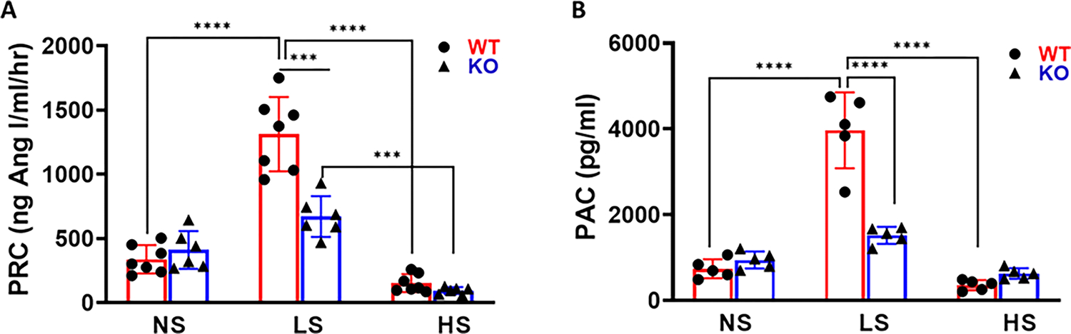
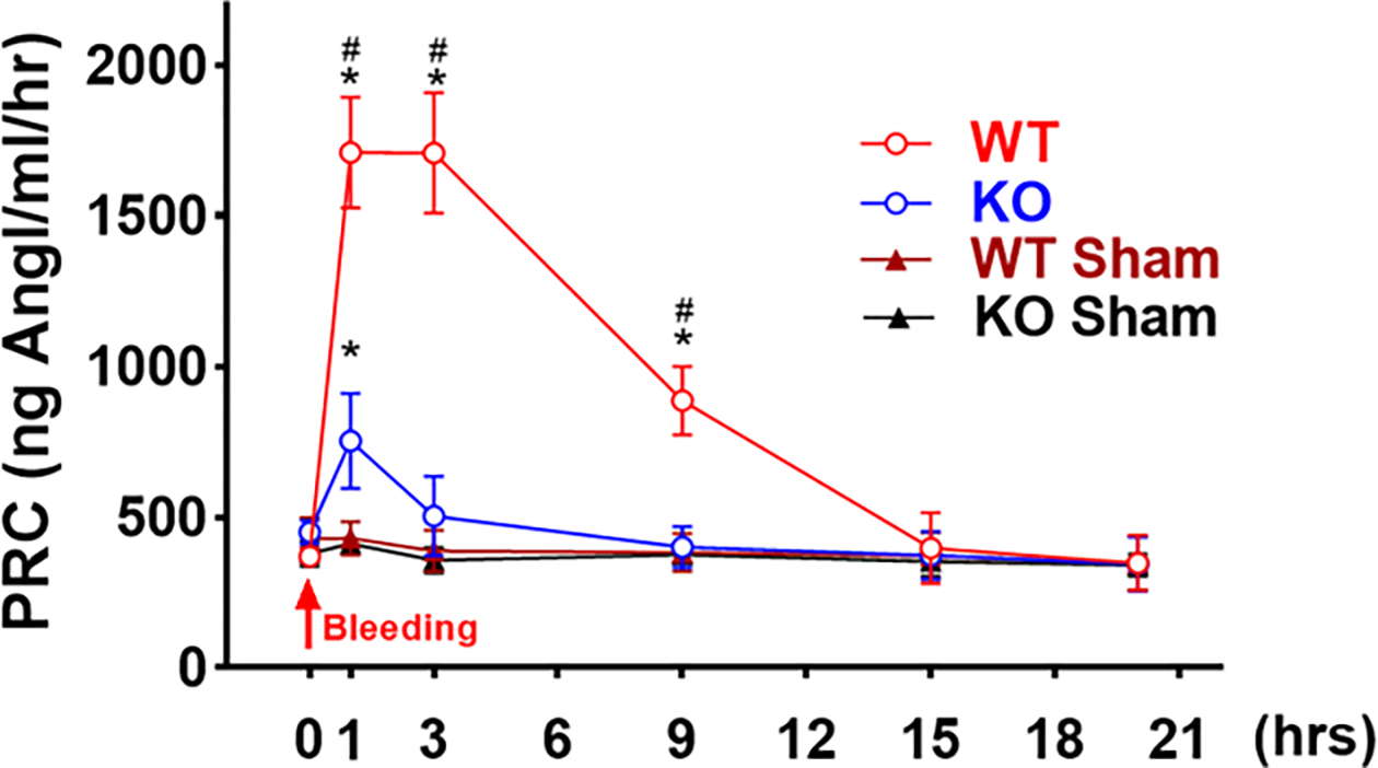
PRCs were 371 ± 95 and 411 ± 68 ng/mL/hr in WT and MD-NOS1KO mice fed a normal sodium diet, respectively. Low salt intake stimulated an increase in the renin release by about 260% in WT mice (PRC = 1364 ± 217 ng/mL/hr, < 0.0001) compared to the PRC under normal salt diet. However, the stimulation was significantly blunted in MD-NOS1KO mice (PRC = 678 ± 104 ng/mL/hr, < 0.001). High salt intake suppressed the PRC to about 61% of the PRC level under a normal salt diet ( < 0.0001). Deletion of macula densa NOS1 further inhibited renin release to 33% of the levels of a normal salt diet. Hemorrhagic shock induced about a 3-fold increase in PRC in WT mice, but only about a 54% increase in the MD-NOS1KO mice ( < 0.0001). The MAP values were substantially greater in WT mice than in MD-NOS1KO mice within the first 6 hours following hemorrhagic shock ( < 0.001). Thus, WT mice showed a much quicker recovery in MAP than MD-NOS1KO mice.
Our study demonstrated that macula densa NOS1 plays an important role in mediating renin release. This mechanism is essential in maintaining blood pressure under hypovolemic situations such as hemorrhagic shock.
关于 (macula densa nitric oxide synthase 1) 对肾素释放的调节作用一直存在争议。本研究旨在进一步阐明 (macula densa) 中 (nitric oxide synthase 1) 在盐挑战和失血性休克时调节肾素释放和血压的作用。
为了研究饮食中不同钠浓度下 (macula densa nitric oxide synthase 1) 在 (macula densa) 中的特定作用,组织 (macula densa-specific) 敲除 NOS1(MD-NOS1KO)和野生型(WT)小鼠分别接受低(0.1%NaCl)和高(1.4%NaCl)盐饮食。通过眼眶后静脉采血 12 mL/kg 体重诱导失血性休克,分别建立包括 MD-NOS1KO 亚组和 WT 亚组在内的两组小鼠。采用无线电遥测系统测量平均动脉压(MAP),采用放射免疫法测量盐饮食和失血性休克实验的血浆肾素浓度(PRC)。
WT 和 MD-NOS1KO 小鼠分别在正常钠饮食和低盐饮食下的 PRC 为 371 ± 95 和 411 ± 68ng/mL/hr。低盐摄入刺激 WT 小鼠的肾素释放增加约 260%(PRC = 1364 ± 217ng/mL/hr, < 0.0001),与正常盐饮食下的 PRC 相比。然而,在 MD-NOS1KO 小鼠中,这种刺激明显减弱(PRC = 678 ± 104ng/mL/hr, < 0.001)。高盐摄入将 PRC 抑制至正常盐饮食下的 61%左右( < 0.0001)。敲除 (macula densa nitric oxide synthase 1) 进一步将肾素释放抑制至正常盐饮食水平的 33%。失血性休克诱导 WT 小鼠的 PRC 增加约 3 倍,但仅在 MD-NOS1KO 小鼠中增加约 54%( < 0.0001)。在失血性休克后最初 6 小时内,WT 小鼠的 MAP 值明显大于 MD-NOS1KO 小鼠( < 0.001)。因此,WT 小鼠比 MD-NOS1KO 小鼠更快地恢复 MAP。
我们的研究表明, (macula densa nitric oxide synthase 1) 在介导肾素释放中起着重要作用。在失血性休克等低血容量情况下,这种机制对于维持血压至关重要。