Department of Radiology, Second Affiliated Hospital of Naval Medical University, Shanghai, China.
Department of Radiology, Xuzhou Medical University, School of Medical Imaging, Xuzhou, China.
Diagn Interv Radiol. 2023 Sep 5;29(5):691-703. doi: 10.4274/dir.2023.232233. Epub 2023 Aug 10.
To assess the quantification accuracy of pulmonary nodules using virtual monoenergetic images (VMIs) derived from spectral-detector computed tomography (CT) under an ultra-low-dose scan protocol.
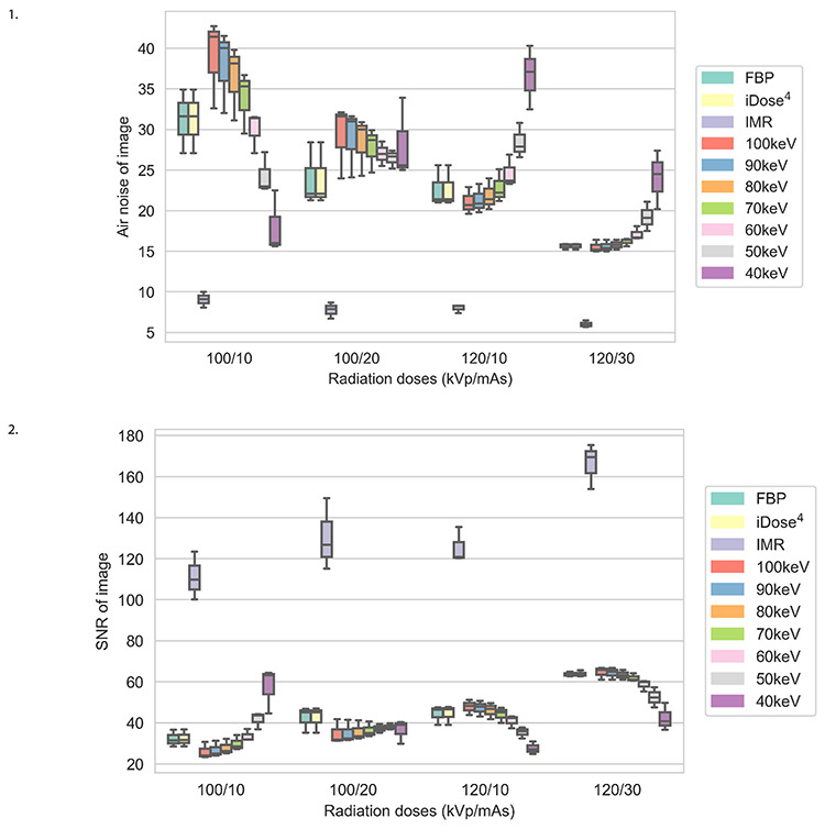
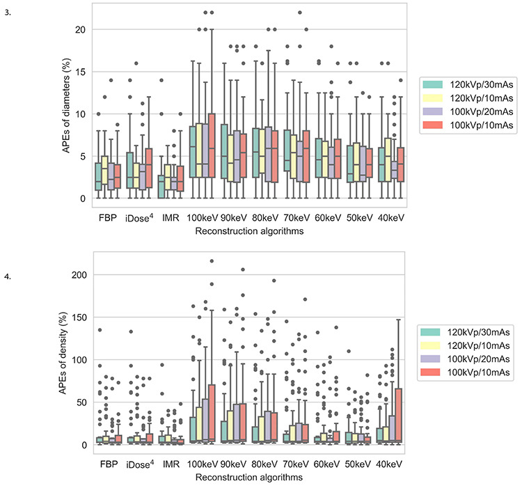
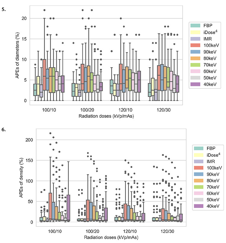
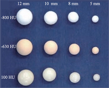
A chest phantom consisting of 12 pulmonary nodules was scanned using spectral-detector CT at 100 kVp/10 mAs, 100 kVp/20 mAs, 120 kVp/10 mAs, and 120 kVp/30 mAs. Each scanning protocol was repeated three times. Each CT scan was reconstructed utilizing filtered back projection, hybrid iterative reconstruction, iterative model reconstruction (IMR), and VMIs of 40-100 keV. The signal-to-noise ratio and air noise of images, absolute differences, and absolute percentage measurement errors (APEs) of the diameter, density, and volume of the four scan protocols and ten reconstruction images were compared.
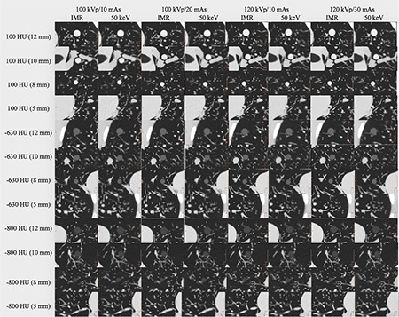
With each fixed reconstruction image, the four scanning protocols exhibited no significant differences in APEs for diameter and density (all > 0.05). Of the four scan protocols and ten reconstruction images, APEs for nodule volume had no significant differences (all > 0.05). At 100 kVp/10 mAs, APEs for density using IMR were the lowest (APE: 6.69), but no significant difference was detected between VMIs at 50 keV (APE: 11.69) and IMR ( = 0.666). In the subgroup analysis, at 100 kVp/10 mAs, there were no significant differences between VMIs at 50 keV and IMR in diameter and density (all > 0.05). The radiation dose at 100 kVp/10 mAs was reduced by 77.8% compared with that at 120 kVp/30 mAs.
Compared with IMR, reconstruction at 100 kVp/10 mAs and 50 keV provides a more accurate quantification of pulmonary nodules, and the radiation dose is reduced by 77.8% compared with that at 120 kVp/30 mAs, demonstrating great potential for ultra-low-dose spectral-detector CT.
评估在超低剂量扫描方案下,使用光谱探测器 CT 生成的虚拟单能量图像(VMIs)对肺结节进行定量准确性。
使用光谱探测器 CT 在 100 kVp/10 mAs、100 kVp/20 mAs、120 kVp/10 mAs 和 120 kVp/30 mAs 下对包含 12 个肺结节的胸部体模进行扫描。每种扫描方案重复 3 次。每个 CT 扫描都使用滤波反投影、混合迭代重建、迭代模型重建(IMR)和 40-100 keV 的 VMIs 进行重建。比较了四种扫描方案和十种重建图像的图像信噪比和空气噪声、直径、密度和体积的绝对差值和绝对百分比测量误差(APE)。
在每种固定的重建图像中,四种扫描方案的直径和密度的 APE 没有显著差异(均>0.05)。在四种扫描方案和十种重建图像中,结节体积的 APE 没有显著差异(均>0.05)。在 100 kVp/10 mAs 下,使用 IMR 的密度的 APE 最低(APE:6.69),但在 50 keV 的 VMIs(APE:11.69)和 IMR(=0.666)之间未检测到显著差异。在亚组分析中,在 100 kVp/10 mAs 下,50 keV 的 VMIs 和 IMR 在直径和密度方面没有显著差异(均>0.05)。与 120 kVp/30 mAs 相比,100 kVp/10 mAs 的辐射剂量降低了 77.8%。
与 IMR 相比,在 100 kVp/10 mAs 和 50 keV 下进行重建可更准确地对肺结节进行定量,并且与 120 kVp/30 mAs 相比,辐射剂量降低了 77.8%,这表明在超低剂量光谱探测器 CT 中具有很大的潜力。