Atlanta Veterans Affairs Medical Center, Decatur, United States.
Department of Orthopaedics, Emory University School of Medicine, Atlanta, United States.
Elife. 2023 Aug 10;12:e63402. doi: 10.7554/eLife.63402.
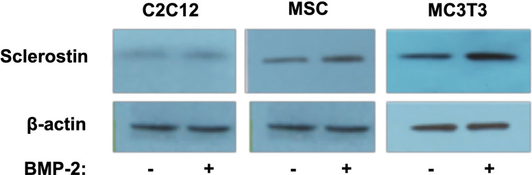
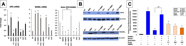
The clinical healing environment after a posterior spinal arthrodesis surgery is one of the most clinically challenging bone-healing environments across all orthopedic interventions due to the absence of a contained space and the need to form de novo bone. Our group has previously reported that sclerostin in expressed locally at high levels throughout a developing spinal fusion. However, the role of sclerostin in controlling bone fusion remains to be established.
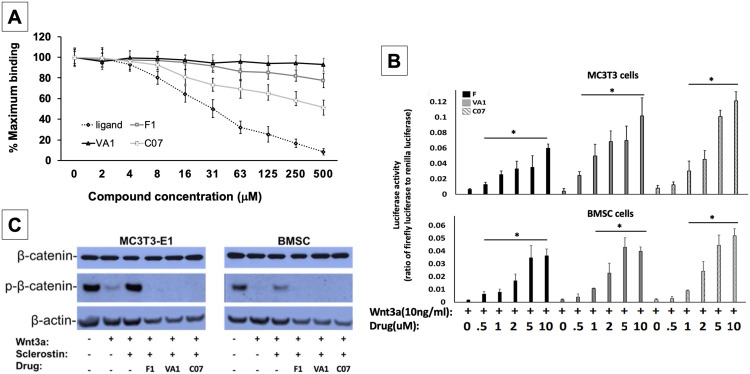
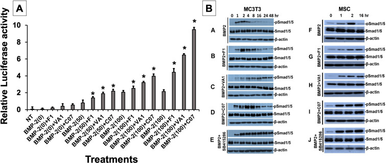
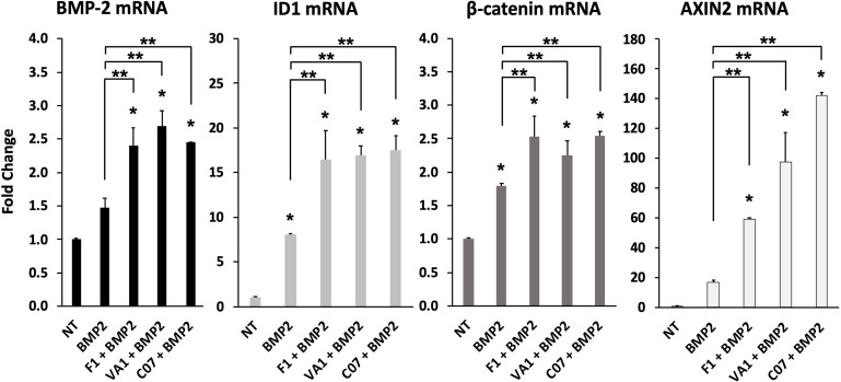
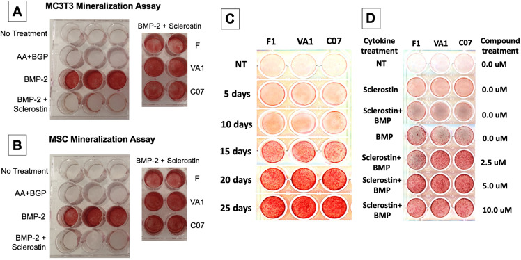
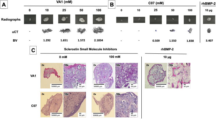
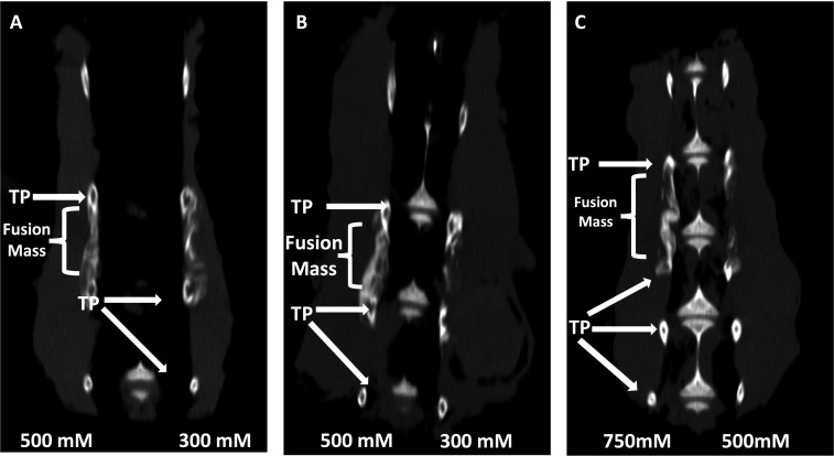
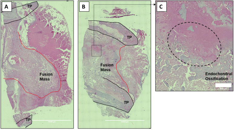
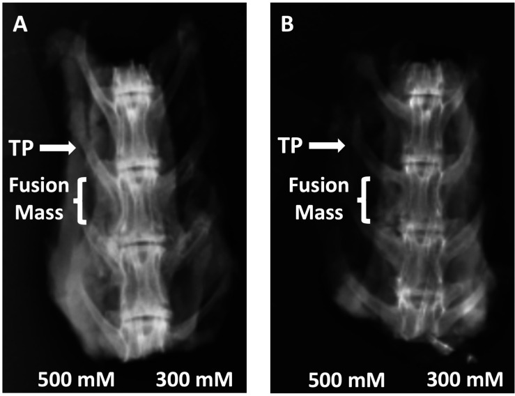
We computationally identified two FDA-approved drugs, as well as a single novel small-molecule drug, for their ability to disrupt the interaction between sclerostin and its receptor, LRP5/6. The drugs were tested in several in vitro biochemical assays using murine MC3T3 and MSCs, assessing their ability to (1) enhance canonical Wnt signaling, (2) promote the accumulation of the active (non-phosphorylated) form of β-catenin, and (3) enhance the intensity and signaling duration of BMP signaling. These drugs were then tested subcutaneously in rats as standalone osteoinductive agents on plain collagen sponges. Finally, the top drug candidates (called VA1 and C07) were tested in a rabbit posterolateral spine fusion model for their ability to achieve a successful fusion at 6 wk.
We show that by controlling GSK3b phosphorylation our three small-molecule inhibitors (SMIs) simultaneously enhance canonical Wnt signaling and potentiate canonical BMP signaling intensity and duration. We also demonstrate that the SMIs produce dose-dependent ectopic mineralization in vivo in rats as well as significantly increase posterolateral spine fusion rates in rabbits in vivo, both as standalone osteogenic drugs and in combination with autologous iliac crest bone graft.
Few if any osteogenic small molecules possess the osteoinductive potency of BMP itself - that is, the ability to form de novo ectopic bone as a standalone agent. Herein, we describe two such SMIs that have this unique ability and were shown to induce de novo bone in a stringent in vivo environment. These SMIs may have the potential to be used in novel, cost-effective bone graft substitutes for either achieving spinal fusion or in the healing of critical-sized fracture defects.
This work was supported by a Veteran Affairs Career Development Award (IK2-BX003845).
后路脊柱融合术后的临床愈合环境是所有骨科介入中最具临床挑战性的骨愈合环境之一,因为它没有封闭的空间,需要形成新骨。我们的研究小组之前报道过,硬化蛋白在正在形成的脊柱融合过程中在局部高水平表达。然而,硬化蛋白在控制骨融合中的作用仍有待确定。
我们通过计算机识别出两种已获得美国食品和药物管理局批准的药物,以及一种新型小分子药物,以评估它们破坏硬化蛋白与其受体 LRP5/6 相互作用的能力。这些药物在使用小鼠 MC3T3 和 MSC 的几种体外生化测定中进行了测试,评估它们(1)增强经典 Wnt 信号传导,(2)促进非磷酸化的β-catenin 的积累,以及(3)增强 BMP 信号的强度和持续时间的能力。然后,这些药物在大鼠中作为单独的成骨诱导剂在普通胶原海绵上进行皮下测试。最后,对顶级候选药物(称为 VA1 和 C07)在兔后路脊柱融合模型中进行了测试,以评估它们在 6 周时实现成功融合的能力。
我们表明,通过控制 GSK3b 磷酸化,我们的三种小分子抑制剂(SMIs)同时增强了经典 Wnt 信号传导,并增强了经典 BMP 信号强度和持续时间。我们还证明,SMIs 在体内产生剂量依赖性异位矿化,并且在体内显著增加兔后路脊柱融合率,作为单独的成骨药物以及与自体髂嵴骨移植物联合使用。
很少有骨形成小分子具有 BMP 本身的成骨潜能——即作为单独的药物形成新的异位骨的能力。在这里,我们描述了两种具有这种独特能力的 SMIs,并证明它们能够在严格的体内环境中诱导新骨形成。这些 SMIs 可能具有作为新型、具有成本效益的骨移植物替代品的潜力,用于实现脊柱融合或治疗临界尺寸骨折缺陷的愈合。
这项工作得到了退伍军人事务部职业发展奖(IK2-BX003845)的支持。