• 文献检索
  • 文档翻译
  • 深度研究
  • 学术资讯
  • Suppr Zotero 插件Zotero 插件
  • 邀请有礼
  • 套餐&价格
  • 历史记录
应用&插件
Suppr Zotero 插件Zotero 插件浏览器插件Mac 客户端Windows 客户端微信小程序
定价
高级版会员购买积分包购买API积分包
服务
文献检索文档翻译深度研究API 文档MCP 服务
关于我们
关于 Suppr公司介绍联系我们用户协议隐私条款
关注我们

Suppr 超能文献

核心技术专利:CN118964589B侵权必究
粤ICP备2023148730 号-1Suppr @ 2026

文献检索

告别复杂PubMed语法,用中文像聊天一样搜索,搜遍4000万医学文献。AI智能推荐,让科研检索更轻松。

立即免费搜索

文件翻译

保留排版,准确专业,支持PDF/Word/PPT等文件格式,支持 12+语言互译。

免费翻译文档

深度研究

AI帮你快速写综述,25分钟生成高质量综述,智能提取关键信息,辅助科研写作。

立即免费体验

68镓前列腺特异性膜抗原正电子发射断层扫描-计算机断层扫描中器官局限性前列腺癌的标准化摄取值及其与前列腺特异性抗原水平和 Gleason 评分的相关性

Standardised Uptake Value in Organ Confined Prostate Cancer in 68-Ga- Prostate-Specific Membrane Antigen Positron Emission Tomography-Computed Tomography Scan and its Correlation with Prostate Specific Antigen Level and Gleason Score.

作者信息

Ali Haider, Rashid-Ul-Amin Syed, Hai Abdul

机构信息

Department of Nuclear Medicine and molecular imaging, Sindh Institute of Urology and Transplantation (S.I.U.T.), Karachi, Pakistan.

出版信息

J Cancer Allied Spec. 2023 Aug 13;9(2):529. doi: 10.37029/jcas.v9i2.519. eCollection 2023.

DOI:10.37029/jcas.v9i2.519
PMID:37575209
原文链接:https://pmc.ncbi.nlm.nih.gov/articles/PMC10405982/
Abstract

INTRODUCTION

A positron emission tomography (PET) scan and a computed tomography (CT) scan are an integral part of oncological imaging and other modalities such as magnetic resonance imaging, CT or bone scintigraphy have some limitations in staging the workup of prostate carcinoma. Combined with tissue-specific markers like prostate-specific membrane antigen (PSMA), positron emitter-based functional imaging results have improved. Our study aimed to determine the Standardised Uptake Value (SUVmax) in prostate adenocarcinoma that is confined to the organ in Ga-68-PSMA PET-CT scans and how it correlates with prostate-specific antigen (PSA) levels and Gleason score (GS).

MATERIALS AND METHODS

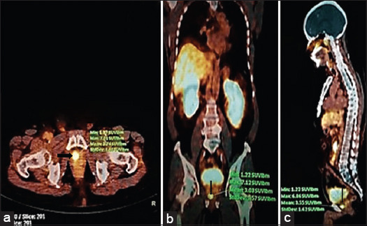
This cross-sectional study was conducted at Sindh Institute of Urology and Transplantation (SIUT), Karachi, and includes subjects referred for a Ga68-PSMA PET-CT scan from September 2017 to January 2022. Histopathologic-proven adenocarcinoma prostate patients with organ-confined disease and PSA levels obtained within 6 weeks before the PSMA-PET-CT scan were included in the study. PET-CT images were semi-quantitatively analysed by measuring SUVmax and the result was interpreted using statistical software SPSS version 22.0.

RESULTS

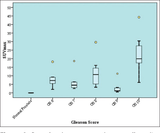
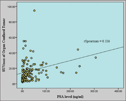
A total of 154 patients were analysed. The mean age of patients was 66.57 ± 8.86 years. The GS of all patients ranges from 6 to 10. The mean and median PSA levels were 32.33 ng/mL (range: 0.004-306.00) and 14.20 ng/mL, respectively. The mean SUVmax of all prostatic lesions was 14.67 ± 12.58 and the median value was 10.76. SUVmax was higher in patients with a PSA level of more than ten than those with a <10. The correlation of SUVmax with PSA and GS showed a significant correlation.

CONCLUSION

The SUVmax of organ-confined prostate cancer correlates well with PSA level and GS Median SUVmax and PSA directly relate to GS.

摘要

引言

正电子发射断层扫描(PET)和计算机断层扫描(CT)是肿瘤成像的重要组成部分,而其他成像方式,如磁共振成像、CT或骨闪烁显像在前列腺癌分期检查中存在一些局限性。结合前列腺特异性膜抗原(PSMA)等组织特异性标志物后,基于正电子发射体的功能成像结果有所改善。我们的研究旨在确定局限于器官内的前列腺腺癌在Ga-68-PSMA PET-CT扫描中的标准化摄取值(SUVmax),以及它与前列腺特异性抗原(PSA)水平和 Gleason评分(GS)的相关性。

材料与方法

本横断面研究在卡拉奇的信德泌尿学与移植研究所(SIUT)进行,纳入了2017年9月至2022年1月期间因Ga68-PSMA PET-CT扫描而转诊的患者。组织病理学证实为前列腺腺癌且疾病局限于器官内,并且在PSMA-PET-CT扫描前6周内获得PSA水平的患者被纳入研究。通过测量SUVmax对PET-CT图像进行半定量分析,并使用统计软件SPSS 22.0对结果进行解读。

结果

共分析了154例患者。患者的平均年龄为66.57±8.86岁。所有患者的GS范围为6至10。PSA的平均水平和中位数分别为32.33 ng/mL(范围:0.004 - 306.00)和14.20 ng/mL。所有前列腺病变的平均SUVmax为14.67±12.58,中位数为10.76。PSA水平大于10的患者的SUVmax高于PSA水平小于10的患者。SUVmax与PSA和GS的相关性显示出显著相关性。

结论

局限于器官内的前列腺癌的SUVmax与PSA水平和GS密切相关。SUVmax中位数和PSA与GS直接相关。

https://cdn.ncbi.nlm.nih.gov/pmc/blobs/2779/10405982/3238010e1f48/JCAS-9-519-g005.jpg
https://cdn.ncbi.nlm.nih.gov/pmc/blobs/2779/10405982/fba87cff908c/JCAS-9-519-g003.jpg
https://cdn.ncbi.nlm.nih.gov/pmc/blobs/2779/10405982/9f0747292f9e/JCAS-9-519-g004.jpg
https://cdn.ncbi.nlm.nih.gov/pmc/blobs/2779/10405982/3238010e1f48/JCAS-9-519-g005.jpg
https://cdn.ncbi.nlm.nih.gov/pmc/blobs/2779/10405982/fba87cff908c/JCAS-9-519-g003.jpg
https://cdn.ncbi.nlm.nih.gov/pmc/blobs/2779/10405982/9f0747292f9e/JCAS-9-519-g004.jpg
https://cdn.ncbi.nlm.nih.gov/pmc/blobs/2779/10405982/3238010e1f48/JCAS-9-519-g005.jpg

相似文献

1
Standardised Uptake Value in Organ Confined Prostate Cancer in 68-Ga- Prostate-Specific Membrane Antigen Positron Emission Tomography-Computed Tomography Scan and its Correlation with Prostate Specific Antigen Level and Gleason Score.68镓前列腺特异性膜抗原正电子发射断层扫描-计算机断层扫描中器官局限性前列腺癌的标准化摄取值及其与前列腺特异性抗原水平和 Gleason 评分的相关性
J Cancer Allied Spec. 2023 Aug 13;9(2):529. doi: 10.37029/jcas.v9i2.519. eCollection 2023.
2
More advantages in detecting bone and soft tissue metastases from prostate cancer using F-PSMA PET/CT.使用F-PSMA PET/CT检测前列腺癌骨和软组织转移方面有更多优势。
Hell J Nucl Med. 2019 Jan-Apr;22(1):6-9. doi: 10.1967/s002449910952. Epub 2019 Mar 7.
3
Ga-PSMA-11 PET/CT in primary staging of prostate cancer: PSA and Gleason score predict the intensity of tracer accumulation in the primary tumour.镓-PSMA-11 PET/CT在前列腺癌初始分期中的应用:前列腺特异抗原(PSA)和 Gleason评分可预测原发肿瘤中示踪剂摄取强度。
Eur J Nucl Med Mol Imaging. 2017 Jun;44(6):941-949. doi: 10.1007/s00259-017-3631-6. Epub 2017 Jan 31.
4
Retrospective correlation of ga-psma uptake with clinical parameters in prostate cancer patients undergoing definitive radiotherapy.回顾性分析前列腺癌患者在接受根治性放疗时 Ga-PSMA 摄取与临床参数的相关性。
Ann Nucl Med. 2020 Jun;34(6):388-396. doi: 10.1007/s12149-020-01462-x. Epub 2020 Mar 27.
5
Diagnostic performance of Gallium-PSMA-11 PET/CT to detect significant prostate cancer and comparison with FEC PET/CT.镓-PSMA-11 PET/CT检测显著前列腺癌的诊断性能及与FEC PET/CT的比较。
Oncotarget. 2017 Nov 14;8(67):111073-111083. doi: 10.18632/oncotarget.22441. eCollection 2017 Dec 19.
6
68Ga-PSMA-11 PET/CT in Newly Diagnosed Prostate Adenocarcinoma.68Ga-PSMA-11 PET/CT 在前列腺腺癌新诊断中的应用。
Clin Nucl Med. 2018 Dec;43(12):e422-e427. doi: 10.1097/RLU.0000000000002289.
7
Incremental value of 68-gallium-prostate-specific membrane antigen positron emission tomography/computed tomography in patients with abnormal prostate-specific antigen and benign transrectal ultrasound biopsy.68镓-前列腺特异性膜抗原正电子发射断层扫描/计算机断层扫描在前列腺特异性抗原异常及经直肠超声引导下活检为良性的患者中的增量价值
Urol Ann. 2018 Apr-Jun;10(2):150-153. doi: 10.4103/UA.UA_55_17.
8
Combined Utility of Ga-Prostate-specific Membrane Antigen Positron Emission Tomography/Computed Tomography and Multiparametric Magnetic Resonance Imaging in Predicting Prostate Biopsy Pathology.前列腺特异性膜抗原正电子发射断层扫描/计算机断层扫描与多参数磁共振成像联合预测前列腺穿刺活检病理。
Eur Urol Oncol. 2022 Jun;5(3):314-320. doi: 10.1016/j.euo.2021.02.006. Epub 2021 Mar 23.
9
Incremental Impact of [ Ga]Ga-PSMA-11 PET/CT in Primary N and M Staging of Prostate Cancer Prior to Curative-Intent Surgery: a Prospective Clinical Trial in Comparison with mpMRI.[68Ga]Ga-PSMA-11 PET/CT 对前列腺癌根治性手术前 N 和 M 分期的增量影响:与 mpMRI 比较的前瞻性临床试验。
Mol Imaging Biol. 2022 Feb;24(1):50-59. doi: 10.1007/s11307-021-01650-9. Epub 2021 Sep 14.
10
(68)Ga-PSMA-11 dynamic PET/CT imaging in biochemical relapse of prostate cancer.(68)镓-PSMA-11动态PET/CT成像在前列腺癌生化复发中的应用
Eur J Nucl Med Mol Imaging. 2016 Jul;43(7):1288-99. doi: 10.1007/s00259-015-3302-4. Epub 2016 Jan 12.

引用本文的文献

1
Semiquantitative Analysis in PET/CT Imaging of Prostate Cancer.前列腺癌PET/CT成像中的半定量分析。
J Clin Med. 2025 May 29;14(11):3828. doi: 10.3390/jcm14113828.

本文引用的文献

1
Determining the diagnostic value of PSMA-PET/CT imaging in patients with persistent high prostate specific antigen levels and negative prostate biopsies.评估 PSMA-PET/CT 成像在前列腺特异性抗原持续升高且前列腺活检阴性患者中的诊断价值。
Urol Oncol. 2022 Feb;40(2):58.e1-58.e7. doi: 10.1016/j.urolonc.2021.07.010. Epub 2021 Aug 14.
2
Establishment and prospective validation of an SUV cutoff value to discriminate clinically significant prostate cancer from benign prostate diseases in patients with suspected prostate cancer by Ga-PSMA PET/CT: a real-world study.应用 Ga-PSMA PET/CT 建立并前瞻性验证 SUV 截断值以鉴别疑似前列腺癌患者中由前列腺特异性膜抗原 PET/CT 检测的临床显著前列腺癌与良性前列腺疾病:一项真实世界研究。
Theranostics. 2021 Jul 25;11(17):8396-8411. doi: 10.7150/thno.58140. eCollection 2021.
3
EAU-EANM-ESTRO-ESUR-SIOG Guidelines on Prostate Cancer-2020 Update. Part 1: Screening, Diagnosis, and Local Treatment with Curative Intent.EAU-EANM-ESTRO-ESUR-SIOG 前列腺癌指南-2020 版更新。第 1 部分:筛查、诊断和以治愈为目的的局部治疗。
Eur Urol. 2021 Feb;79(2):243-262. doi: 10.1016/j.eururo.2020.09.042. Epub 2020 Nov 7.
4
Diagnostic Accuracy of Ga-PSMA PET/CT for Initial Detection in Patients With Suspected Prostate Cancer: A Systematic Review and Meta-Analysis.Ga-PSMA PET/CT 对疑似前列腺癌患者初始检测的诊断准确性:系统评价和荟萃分析。
AJR Am J Roentgenol. 2021 Mar;216(3):599-607. doi: 10.2214/AJR.20.23912. Epub 2021 Jan 21.
5
Ga-PSMA PET/CT targeted biopsy for the diagnosis of clinically significant prostate cancer compared with transrectal ultrasound guided biopsy: a prospective randomized single-centre study.镓-PSMA PET/CT 靶向活检与经直肠超声引导活检诊断临床显著前列腺癌的比较:一项前瞻性随机单中心研究。
Eur J Nucl Med Mol Imaging. 2021 Feb;48(2):483-492. doi: 10.1007/s00259-020-04863-2. Epub 2020 Jul 30.
6
Prostate Magnetic Resonance Imaging, with or Without Magnetic Resonance Imaging-targeted Biopsy, and Systematic Biopsy for Detecting Prostate Cancer: A Cochrane Systematic Review and Meta-analysis.前列腺磁共振成像联合或不联合磁共振成像靶向活检与系统活检用于前列腺癌检测的效果:一项 Cochrane 系统评价和荟萃分析。
Eur Urol. 2020 Jan;77(1):78-94. doi: 10.1016/j.eururo.2019.06.023. Epub 2019 Jul 18.
7
Can SUVmax values of Ga-68-PSMA PET/CT scan predict the clinically significant prostate cancer?Ga-68-PSMA PET/CT扫描的SUVmax值能否预测具有临床意义的前列腺癌?
Nucl Med Commun. 2019 Jan;40(1):86-91. doi: 10.1097/MNM.0000000000000942.
8
Re: MRI-Targeted or Standard Biopsy for Prostate-Cancer Diagnosis.关于:MRI靶向活检还是标准活检用于前列腺癌诊断
J Urol. 2018 Oct;200(4):697-699. doi: 10.1016/j.juro.2018.07.026. Epub 2018 Jul 17.
9
Prostate Imaging-Reporting and Data System Steering Committee: PI-RADS v2 Status Update and Future Directions.前列腺影像报告和数据系统指导委员会:PI-RADS v2 现状更新及未来方向。
Eur Urol. 2019 Mar;75(3):385-396. doi: 10.1016/j.eururo.2018.05.035. Epub 2018 Jun 13.
10
Negative Multiparametric Magnetic Resonance Imaging for Prostate Cancer: What's Next?前列腺癌的负多参数磁共振成像:下一步是什么?
Eur Urol. 2018 Jul;74(1):48-54. doi: 10.1016/j.eururo.2018.03.007. Epub 2018 Mar 19.