Radiology Department, Second Affiliated Hospital of Zhejiang Chinese Medical University, Hangzhou, 310005, China.
Department of Radiology, the People's Hospital of Jianyang City, Chengdu, 641499, China.
J Digit Imaging. 2023 Dec;36(6):2554-2566. doi: 10.1007/s10278-023-00888-9. Epub 2023 Aug 14.
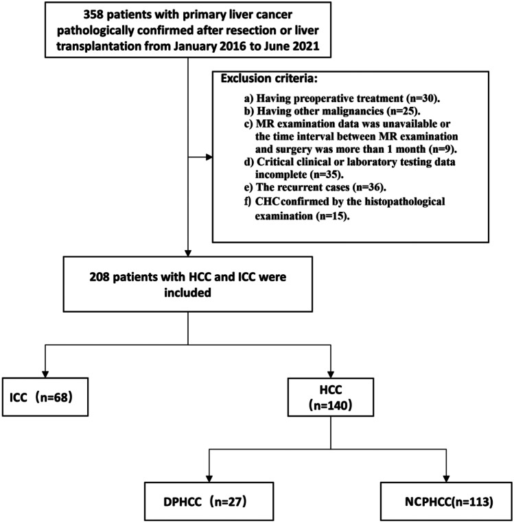
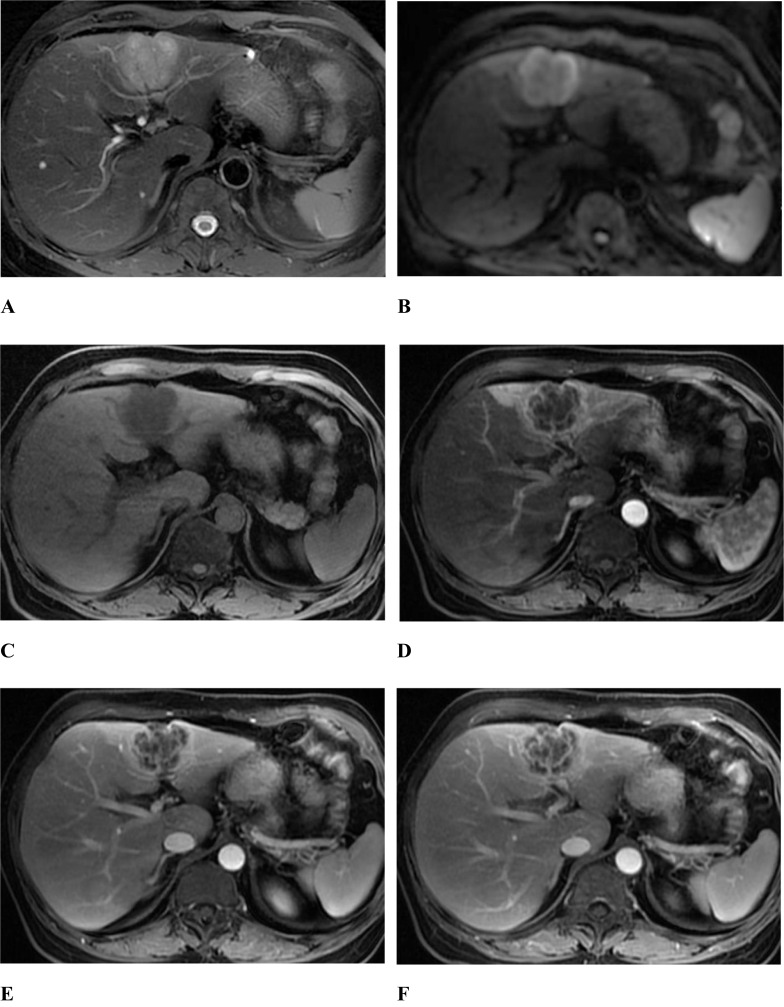
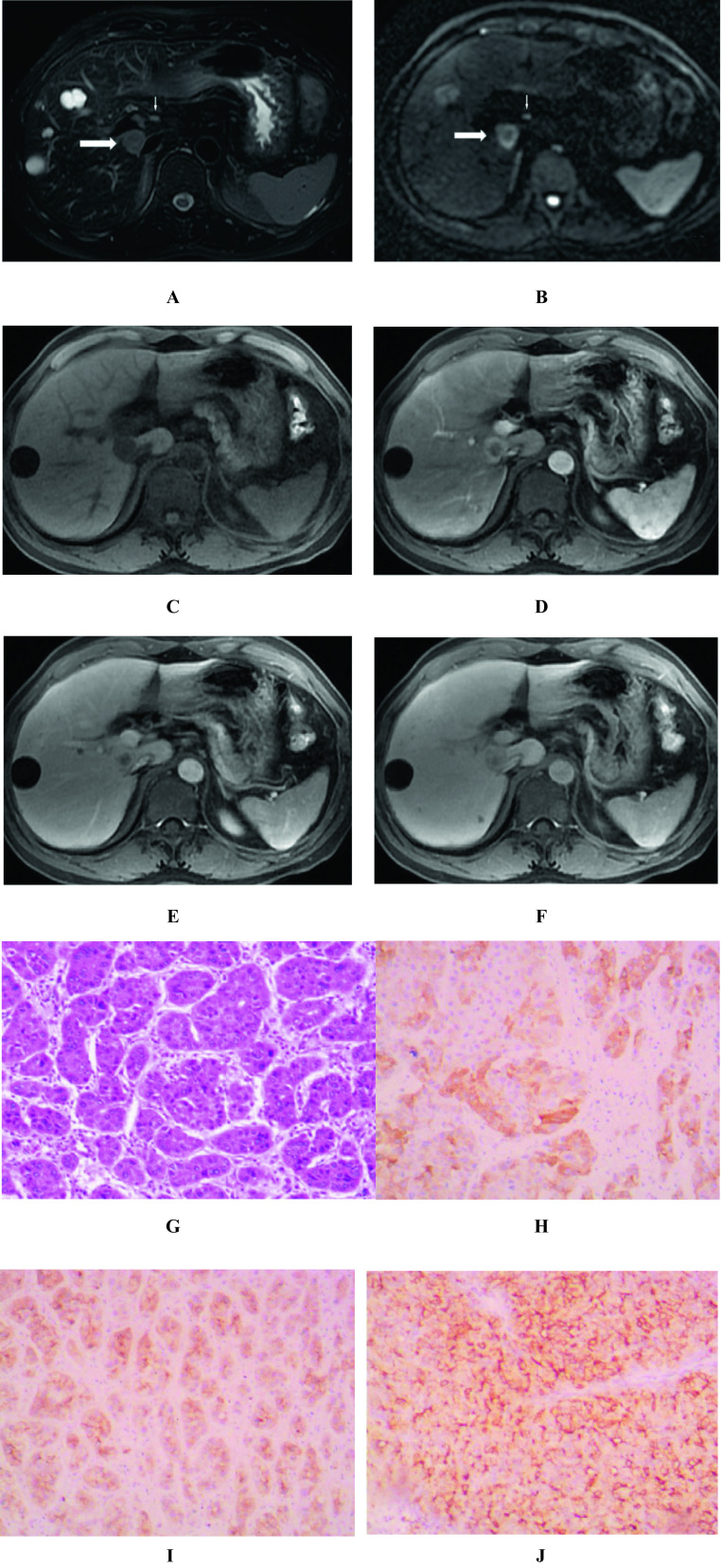
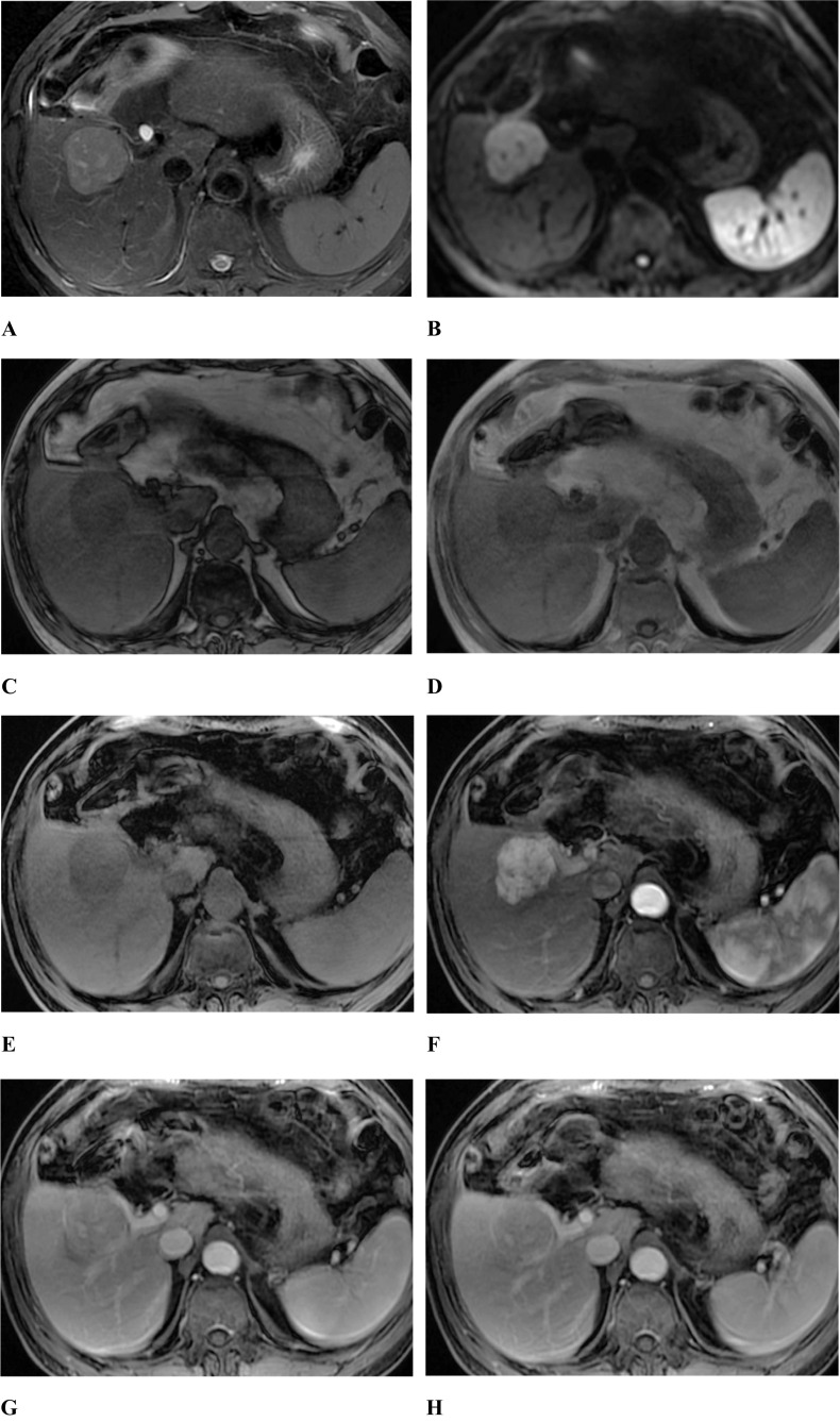
This study aimed to explore the magnetic resonance imaging (MRI) features of dual-phenotype hepatocellular carcinoma (DPHCC) and their diagnostic value.The data of 208 patients with primary liver cancer were retrospectively analysed between January 2016 and June 2021. Based on the pathological diagnostic criteria, 27 patients were classified into the DPHCC group, 113 patients into the noncholangiocyte-phenotype hepatocellular carcinoma (NCPHCC) group, and 68 patients with intrahepatic cholangiocarcinoma (ICC) were classified into the ICC group. Two abdominal radiologists reviewed the preoperative MRI features by a double-blind method. The MRI features and key laboratory and clinical indicators were compared between the groups. The potentially valuable MRI features and key laboratory and clinical characteristics for predicting DPHCC were identified by univariate and multivariate analyses, and the odds ratios (ORs) were recorded. In multivariate analysis, tumour without capsule (P = 0.046, OR = 9.777), dynamic persistent enhancement (P = 0.006, OR = 46.941), and targetoid appearance on diffusion-weighted imaging (DWI) (P = 0.021, OR = 30.566) were independently significant factors in the detection of DPHCC compared to NCPHCC. Serum alpha-fetoprotein (AFP) > 20 µg/L (P = 0.036, OR = 67.097) and prevalence of hepatitis B virus (HBV) infection (P = 0.020, OR = 153.633) were independent significant factors in predicting DPHCC compared to ICC. The differences in other tumour marker levels and imaging features between the groups were not significant. In MR enhanced and diffusion imaging, tumour without capsule, persistent enhancement and DWI targetoid findings, combined with AFP > 20 µg/L and HBV infection-positive laboratory results, can help to diagnose DPHCC and differentiate it from NCPHCC and ICC. These results suggest that clinical, laboratory and MRI features should be integrated to construct an AI diagnostic model for DPHCC.
本研究旨在探讨双表型肝细胞癌(DPHCC)的磁共振成像(MRI)特征及其诊断价值。回顾性分析 2016 年 1 月至 2021 年 6 月间收治的 208 例原发性肝癌患者资料,根据病理诊断标准将 27 例患者分为 DPHCC 组,113 例患者分为非胆管细胞型肝细胞癌(NCPHCC)组,68 例患者分为肝内胆管细胞癌(ICC)组。两名腹部放射科医生采用双盲法对术前 MRI 特征进行回顾性分析。比较各组间 MRI 特征及实验室和临床指标。采用单因素和多因素分析确定预测 DPHCC 的有价值的 MRI 特征及实验室和临床特征,记录比值比(OR)。多因素分析显示,肿瘤无包膜(P=0.046,OR=9.777)、动态持续强化(P=0.006,OR=46.941)和 DWI 上靶样表现(P=0.021,OR=30.566)是与 NCPHCC 相比,检测 DPHCC 的独立显著因素。血清甲胎蛋白(AFP)>20μg/L(P=0.036,OR=67.097)和乙型肝炎病毒(HBV)感染(P=0.020,OR=153.633)是与 ICC 相比预测 DPHCC 的独立显著因素。各组间其他肿瘤标志物水平和影像学特征差异无统计学意义。MR 增强和扩散成像中,肿瘤无包膜、持续强化和 DWI 靶样表现,结合 AFP>20μg/L 和 HBV 感染阳性实验室结果,有助于诊断 DPHCC,并与 NCPHCC 和 ICC 相鉴别。这些结果提示,临床、实验室和 MRI 特征应整合构建 DPHCC 的人工智能诊断模型。