Department of Neurosurgery, Affiliated Hospital of Nantong University, Medical School of Nantong University, Nantong, China.
Research Center of Clinical Medicine, Affiliated Hospital of Nantong University, Nantong, China.
CNS Neurosci Ther. 2024 Feb;30(2):e14412. doi: 10.1111/cns.14412. Epub 2023 Aug 17.
The current evidence demonstrates that mesenchymal stem cells (MSCs) hold therapeutic potential for ischemic stroke. However, it remains unclear how changes in the secretion of MSC cytokines following the overexpression of heme oxygenase-1 (HO-1) impact excessive inflammatory activation in a mouse ischemic stroke model. This study investigated this aspect and provided further insights.
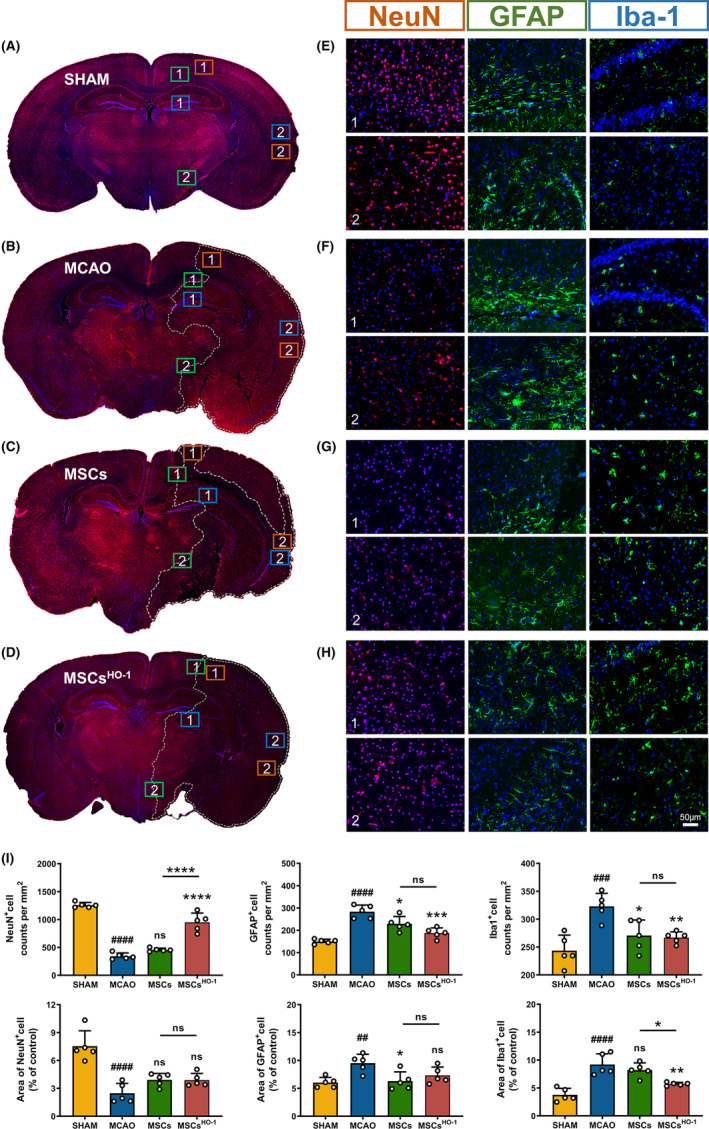
The middle cerebral artery occlusion (MCAO) mouse model was established, and subsequent injections of MSC, MSC , or PBS solutions of equal volume were administered via the mice's tail vein. Histopathological analysis was conducted on Days 3 and 28 post-MCAO to observe morphological changes in brain slices. mRNA expression levels of various factors, including IL-1β, IL-6, IL-17, TNF-α, IL-1Ra, IL-4, IL-10, TGF-β, were quantified. The effects of MSC treatment on neurons, microglia, and astrocytes were observed using immunofluorescence after transplantation. The polarization direction of macrophages/microglia was also detected using flow cytometry.
The results showed that the expression of anti-inflammatory factors in the MSC group increased while that of pro-inflammatory factors decreased. Small animal fluorescence studies and immunofluorescence assays showed that the homing function of MSCs was unaffected, leading to a substantial accumulation of MSCs in the cerebral ischemic region within 24 h. Neurons were less damaged, activation and proliferation of microglia were reduced, and polarization of microglia to the M2 type increased after MSC transplantation. Furthermore, after transplantation of MSCs , the mortality of mice decreased, and motor function improved significantly.
The findings indicate that MSCs overexpressing HO-1 exhibited significant therapeutic effects against hyper-inflammatory injury after stroke in mice, ultimately promoting recovery after ischemic stroke.
目前的证据表明间充质干细胞(MSCs)在缺血性中风中具有治疗潜力。然而,尚不清楚过表达血红素加氧酶-1(HO-1)后 MSC 细胞因子分泌的变化如何影响小鼠缺血性中风模型中的过度炎症激活。本研究对此进行了探讨,并提供了进一步的见解。
建立大脑中动脉闭塞(MCAO)小鼠模型,随后通过小鼠尾静脉注射 MSC、MSC 或等量 PBS 溶液。在 MCAO 后第 3 天和第 28 天进行脑切片的组织病理学分析,观察形态学变化。定量检测各种因子(包括 IL-1β、IL-6、IL-17、TNF-α、IL-1Ra、IL-4、IL-10、TGF-β)的 mRNA 表达水平。移植后使用免疫荧光观察 MSC 治疗对神经元、小胶质细胞和星形胶质细胞的影响。还使用流式细胞术检测巨噬细胞/小胶质细胞的极化方向。
结果表明,MSC 组抗炎因子的表达增加,促炎因子的表达减少。小动物荧光研究和免疫荧光分析表明,MSCs 的归巢功能不受影响,导致 MSCs 在脑缺血区域内 24 小时内大量聚集。神经元损伤减轻,小胶质细胞的激活和增殖减少,MSC 移植后小胶质细胞向 M2 型极化增加。此外,MSC 移植后,小鼠死亡率降低,运动功能明显改善。
这些发现表明,过表达 HO-1 的 MSC 对小鼠中风后过度炎症损伤具有显著的治疗作用,最终促进缺血性中风后的恢复。