From the Institute of Ophthalmology (S.K.W., D.J.W., R.R.S., Y.Z., P.J.F., K.B., A.P.K., P.J.P., J.S.R., A.P., P.A.K.), University College London; NIHR Biomedical Research Centre at Moorfields Eye Hospital and UCL Institute of Ophthalmology (S.K.W., D.R.-B., D.J.W., R.R.S., Y.Z., E.K., P.J.F., K.B., A.P.K., P.J.P., J.S.R., A.K.D., A.P., P.A.K.), London, United Kingdom; Biomedical Engineering Department (D.R.-B., E.K., U.A., M.B.), Faculty of Engineering (MU-ENG), Mondragon Unibertsitatea, Spain; Great Ormond Street Institute of Child Health (M.C.-B., J.S.R.), and Centre for Medical Image Computing (D.J.W., R.R.S., Y.Z.), Department of Computer Science, University College London; NeuroMetrology Lab (S.P., C.A.A.), Nuffield Department of Clinical Neurosciences, University of Oxford; Dementia Research Centre (R.S.W.), University College London, United Kingdom; Department of Molecular Medicine (E.J.T.), Scripps Research, La Jolla, CA; Byers Eye Institute (E.K.), Stanford University, Palo Alto, CA; Biocruces Bizkaia Health Research Institute (I.G.), Barakaldo; IKERBASQUE: The Basque Foundation for Science (I.G.), Bilbao, Spain; Department of Clinical and Movement Neurosciences (A.H.V.S.), UCL Queen Square Institute of Neurology; Great Ormond Street Hospital NHS Foundation Trust (J.S.R.); Ulverscroft Vision Research Group (J.S.R.), University College London; NIHR Biomedical Research Centre at UCL Great Ormond Street Institute of Child Health and Great Ormond Street Hospital (J.S.R.), London; University of Birmingham (A.K.D.); University Hospitals Birmingham NHS Foundation Trust (A.K.D.); NIHR Birmingham Biomedical Research Centre (A.K.D.), University of Birmingham; and Queen Square Institute of Neurology (A.P.), University College London, United Kingdom.
Neurology. 2023 Oct 17;101(16):e1581-e1593. doi: 10.1212/WNL.0000000000207727. Epub 2023 Aug 21.
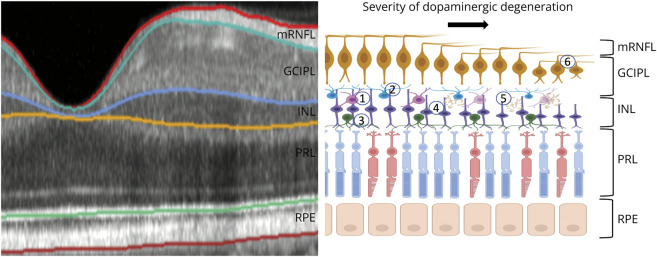
Cadaveric studies have shown disease-related neurodegeneration and other morphological abnormalities in the retina of individuals with Parkinson disease (PD); however, it remains unclear whether this can be reliably detected with in vivo imaging. We investigated inner retinal anatomy, measured using optical coherence tomography (OCT), in prevalent PD and subsequently assessed the association of these markers with the development of PD using a prospective research cohort.
This cross-sectional analysis used data from 2 studies. For the detection of retinal markers in prevalent PD, we used data from AlzEye, a retrospective cohort of 154,830 patients aged 40 years and older attending secondary care ophthalmic hospitals in London, United Kingdom, between 2008 and 2018. For the evaluation of retinal markers in incident PD, we used data from UK Biobank, a prospective population-based cohort where 67,311 volunteers aged 40-69 years were recruited between 2006 and 2010 and underwent retinal imaging. Macular retinal nerve fiber layer (mRNFL), ganglion cell-inner plexiform layer (GCIPL), and inner nuclear layer (INL) thicknesses were extracted from fovea-centered OCT. Linear mixed-effects models were fitted to examine the association between prevalent PD and retinal thicknesses. Hazard ratios for the association between time to PD diagnosis and retinal thicknesses were estimated using frailty models.
Within the AlzEye cohort, there were 700 individuals with prevalent PD and 105,770 controls (mean age 65.5 ± 13.5 years, 51.7% female). Individuals with prevalent PD had thinner GCIPL (-2.12 μm, 95% CI -3.17 to -1.07, = 8.2 × 10) and INL (-0.99 μm, 95% CI -1.52 to -0.47, = 2.1 × 10). The UK Biobank included 50,405 participants (mean age 56.1 ± 8.2 years, 54.7% female), of whom 53 developed PD at a mean of 2,653 ± 851 days. Thinner GCIPL (hazard ratio [HR] 0.62 per SD increase, 95% CI 0.46-0.84, = 0.002) and thinner INL (HR 0.70, 95% CI 0.51-0.96, = 0.026) were also associated with incident PD.
Individuals with PD have reduced thickness of the INL and GCIPL of the retina. Involvement of these layers several years before clinical presentation highlight a potential role for retinal imaging for at-risk stratification of PD.
尸检研究表明,帕金森病(PD)患者的视网膜存在与疾病相关的神经退行性变和其他形态异常;然而,目前尚不清楚是否可以通过活体成像可靠地检测到这些变化。我们使用光学相干断层扫描(OCT)检测了内视网膜解剖结构,并在现患 PD 患者中进行了评估,随后使用前瞻性研究队列评估了这些标志物与 PD 发展的相关性。
本横断面分析使用了两项研究的数据。为了检测现患 PD 中的视网膜标志物,我们使用了来自 AlzEye 的数据,这是一项回顾性队列研究,纳入了 2008 年至 2018 年间在英国伦敦二级保健眼科医院就诊的 154830 名 40 岁及以上的患者。为了评估在新发 PD 中视网膜标志物的情况,我们使用了 UK Biobank 的数据,这是一项前瞻性的基于人群的队列研究,其中 67311 名 40-69 岁的志愿者于 2006 年至 2010 年期间招募,并进行了视网膜成像。从以黄斑为中心的 OCT 中提取黄斑视网膜神经纤维层(mRNFL)、神经节细胞-内丛状层(GCIPL)和内核层(INL)的厚度。使用线性混合效应模型来检验现患 PD 与视网膜厚度之间的关联。使用脆弱性模型估计视网膜厚度与 PD 诊断时间之间的关联的风险比。
在 AlzEye 队列中,有 700 名现患 PD 患者和 105770 名对照者(平均年龄 65.5 ± 13.5 岁,51.7%为女性)。现患 PD 患者的 GCIPL(-2.12 μm,95%CI-3.17 至-1.07, = 8.2×10)和 INL(-0.99 μm,95%CI-1.52 至-0.47, = 2.1×10)较薄。UK Biobank 纳入了 50405 名参与者(平均年龄 56.1 ± 8.2 岁,54.7%为女性),其中 53 名在平均 2653 ± 851 天后发展为 PD。GCIPL 变薄(每标准差增加 HR 0.62,95%CI 0.46-0.84, = 0.002)和 INL 变薄(HR 0.70,95%CI 0.51-0.96, = 0.026)也与新发 PD 相关。
PD 患者的视网膜内核层和神经节细胞-内丛状层厚度减少。这些层在临床症状出现前几年的受累情况提示视网膜成像在 PD 的高危分层中具有潜在作用。