• 文献检索
  • 文档翻译
  • 深度研究
  • 学术资讯
  • Suppr Zotero 插件Zotero 插件
  • 邀请有礼
  • 套餐&价格
  • 历史记录
应用&插件
Suppr Zotero 插件Zotero 插件浏览器插件Mac 客户端Windows 客户端微信小程序
定价
高级版会员购买积分包购买API积分包
服务
文献检索文档翻译深度研究API 文档MCP 服务
关于我们
关于 Suppr公司介绍联系我们用户协议隐私条款
关注我们

Suppr 超能文献

核心技术专利:CN118964589B侵权必究
粤ICP备2023148730 号-1Suppr @ 2026

文献检索

告别复杂PubMed语法,用中文像聊天一样搜索,搜遍4000万医学文献。AI智能推荐,让科研检索更轻松。

立即免费搜索

文件翻译

保留排版,准确专业,支持PDF/Word/PPT等文件格式,支持 12+语言互译。

免费翻译文档

深度研究

AI帮你快速写综述,25分钟生成高质量综述,智能提取关键信息,辅助科研写作。

立即免费体验

澳大利亚肺鱼生长皮肤中某些表皮蛋白和糖蛋白的免疫定位()。 (括号部分原文缺失内容,翻译时保留原样)

Immunolocalization of Some Epidermal Proteins and Glycoproteins in the Growing Skin of the Australian Lungfish ().

作者信息

Alibardi Lorenzo

机构信息

Comparative Histolab Padova, 35100 Padova, Italy.

Department of Biology, University of Bologna, 40126 Bologna, Italy.

出版信息

J Dev Biol. 2023 Aug 14;11(3):35. doi: 10.3390/jdb11030035.

DOI:10.3390/jdb11030035
PMID:37606491
原文链接:https://pmc.ncbi.nlm.nih.gov/articles/PMC10443291/
Abstract

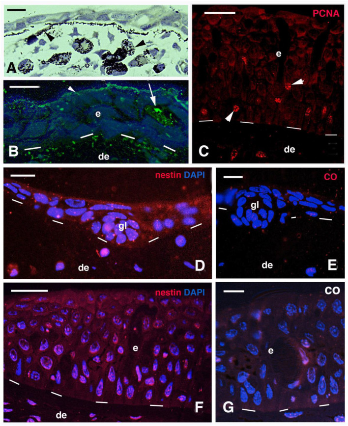
Here we report the immunolocalization of mucin, nestin, elastin and three glycoproteins involved in tissue mineralization in small and large juveniles of . Both small and larger juvenile epidermis are mucogenic and contain a diffuse immunolabeling for nestin. Sparse PCNA-labeled cells, indicating proliferation, are found in basal and suprabasal epidermal layers. No scales are formed in small juveniles but are present in a 5 cm long juvenile and in larger juveniles. Elastin and a mineralizing matrix are localized underneath the basement membrane of the tail epidermis where lepidotriches are forming. The latter appears as "circular bodies" in cross sections and are made of elongated cells surrounding a central amorphous area containing collagen and elastin-like proteins that undergo calcification as evidenced using the von Kossa staining. However, the first calcification sites are the coniform teeth of the small juveniles of 2-3 cm in length. In the superficial dermis of juveniles (16-26 cm in length) where scales are formed, the spinulated outer bony layer (squamulin) of the elasmoid scales contains osteonectin, alkaline phosphatase, osteopontin, and calcium deposits that are instead absent in the underlying layer of elasmodin. In particular, these glycoproteins are localized along the scale margin in juveniles where scales grow, as indicated by the presence of PCNA-labeled cells (proliferating). These observations suggest a continuous deposition of new bone during the growth of the scales, possibly under the action of these mineralizing glycoproteins, like in the endoskeleton of terrestrial vertebrates.

摘要

在此,我们报告了在[具体物种]大小不同的幼年个体中粘蛋白、巢蛋白、弹性蛋白以及三种参与组织矿化的糖蛋白的免疫定位情况。大小不同的幼年个体表皮均具有粘蛋白生成能力,且对巢蛋白呈现弥漫性免疫标记。在基底和基底上层表皮层中发现了稀疏的增殖细胞核抗原(PCNA)标记细胞,表明存在细胞增殖。小的幼年个体未形成鳞片,但在体长5厘米的幼年个体及更大的幼年个体中存在鳞片。弹性蛋白和矿化基质定位于尾表皮基底膜下方,此处正在形成鳞片。后者在横切面上呈现为“圆形体”,由围绕中央无定形区域的细长细胞组成,该区域含有胶原蛋白和类弹性蛋白,经冯·科萨染色证实会发生钙化。然而,最早的钙化部位是体长2 - 3厘米的小幼年个体的锥形齿。在形成鳞片的幼年个体(体长16 - 26厘米)的浅表真皮中,硬鳞的具刺状外层骨层(鳞质)含有骨连接蛋白、碱性磷酸酶、骨桥蛋白和钙沉积物,而在其下方的弹性蛋白层中则不存在这些物质。特别是,这些糖蛋白定位于鳞片生长的幼年个体的鳞片边缘,增殖细胞核抗原(PCNA)标记细胞(正在增殖)的存在表明了这一点。这些观察结果表明,在鳞片生长过程中可能在这些矿化糖蛋白的作用下持续有新骨沉积,类似于陆地脊椎动物的内骨骼。

https://cdn.ncbi.nlm.nih.gov/pmc/blobs/ca94/10443291/9507ca093d46/jdb-11-00035-g007.jpg
https://cdn.ncbi.nlm.nih.gov/pmc/blobs/ca94/10443291/68cc9dceff38/jdb-11-00035-g001.jpg
https://cdn.ncbi.nlm.nih.gov/pmc/blobs/ca94/10443291/f35a351e9ba5/jdb-11-00035-g002.jpg
https://cdn.ncbi.nlm.nih.gov/pmc/blobs/ca94/10443291/59d915e0c0a4/jdb-11-00035-g003.jpg
https://cdn.ncbi.nlm.nih.gov/pmc/blobs/ca94/10443291/cd314e1313e2/jdb-11-00035-g004.jpg
https://cdn.ncbi.nlm.nih.gov/pmc/blobs/ca94/10443291/7f35065d532c/jdb-11-00035-g005.jpg
https://cdn.ncbi.nlm.nih.gov/pmc/blobs/ca94/10443291/95f86ad0a7e9/jdb-11-00035-g006.jpg
https://cdn.ncbi.nlm.nih.gov/pmc/blobs/ca94/10443291/9507ca093d46/jdb-11-00035-g007.jpg

相似文献

1
Immunolocalization of Some Epidermal Proteins and Glycoproteins in the Growing Skin of the Australian Lungfish ().澳大利亚肺鱼生长皮肤中某些表皮蛋白和糖蛋白的免疫定位()。 (括号部分原文缺失内容,翻译时保留原样)
J Dev Biol. 2023 Aug 14;11(3):35. doi: 10.3390/jdb11030035.
2
Scale structure in the Australian lungfish, Neoceratodus forsteri (Osteichthyes: Dipnoi).澳大利亚肺鱼(新角齿鱼属福氏新角齿鱼,硬骨鱼纲:角齿鱼目)的鳞片结构
J Morphol. 2015 Oct;276(10):1137-45. doi: 10.1002/jmor.20399. Epub 2015 Jul 21.
3
Formation and structure of scales in the Australian lungfish, Neoceratodus forsteri (Osteichthyes: Dipnoi).澳大利亚肺鱼(Neoceratodus forsteri)(硬骨鱼纲:角齿鱼目)鳞片的形成与结构
J Morphol. 2012 May;273(5):530-40. doi: 10.1002/jmor.11039. Epub 2011 Dec 21.
4
Skin structure in the snout of the Australian lungfish, Neoceratodus forsteri (Osteichthyes: Dipnoi).澳大利亚肺鱼(新角齿鱼属,福氏新角齿鱼,硬骨鱼纲:肺鱼目)吻部的皮肤结构。
Tissue Cell. 2014 Oct;46(5):397-408. doi: 10.1016/j.tice.2014.07.004. Epub 2014 Aug 11.
5
Visual ecology of the Australian lungfish (Neoceratodus forsteri).澳大利亚肺鱼(新角齿鱼属福氏新角齿鱼)的视觉生态学
BMC Ecol. 2008 Dec 18;8:21. doi: 10.1186/1472-6785-8-21.
6
Ultrastructure of the integumental melanophores of the Australian lungfish, Neoceratodus forsteri.澳大利亚肺鱼(Neoceratodus forsteri)体表黑素细胞的超微结构
Cell Tissue Res. 1975;158(3):363-73. doi: 10.1007/BF00223833.
7
Role of epidermal cilia in development of the Australian lungfish, Neoceratodus forsteri (Osteichthyes: Dipnoi).表皮纤毛在澳大利亚肺鱼(Neoceratodus forsteri)(硬骨鱼纲:角齿鱼目)发育中的作用。
J Morphol. 1996 May;228(2):203-221. doi: 10.1002/(SICI)1097-4687(199605)228:2<203::AID-JMOR9>3.0.CO;2-5.
8
Keratinization of the epidermis of the Australian lungfish Neoceratodus forsteri (dipnoi).
J Morphol. 2003 Apr;256(1):13-22. doi: 10.1002/jmor.10073.
9
Comparative pelvic development of the axolotl (Ambystoma mexicanum) and the Australian lungfish (Neoceratodus forsteri): conservation and innovation across the fish-tetrapod transition.比较蝾螈(Ambystoma mexicanum)和澳大利亚肺鱼(Neoceratodus forsteri)的骨盆发育:鱼类-四足动物过渡中的保护和创新。
Evodevo. 2013 Jan 23;4(1):3. doi: 10.1186/2041-9139-4-3.
10
Cartilage, bone, and intermandibular connective tissue in the Australian lungfish, Neoceratodus forsteri (Osteichthyes: Dipnoi).澳大利亚肺鱼(新角齿鱼属福氏新角齿鱼,硬骨鱼纲:角齿鱼目)的软骨、骨骼和下颌间结缔组织。
J Morphol. 2013 Oct;274(10):1085-9. doi: 10.1002/jmor.20164. Epub 2013 Jun 26.

本文引用的文献

1
Comparison of vertebrate skin structure at class level: A review.比较类水平的脊椎动物皮肤结构:综述。
Anat Rec (Hoboken). 2022 Dec;305(12):3543-3608. doi: 10.1002/ar.24908. Epub 2022 Mar 14.
2
Vertebrate keratinization evolved into cornification mainly due to transglutaminase and sulfhydryl oxidase activities on epidermal proteins: An immunohistochemical survey.脊椎动物角化作用演变为角质化主要是由于表皮蛋白中转谷氨酰胺酶和巯基氧化酶的活性:免疫组织化学调查。
Anat Rec (Hoboken). 2022 Feb;305(2):333-358. doi: 10.1002/ar.24705. Epub 2021 Jul 29.
3
Giant lungfish genome elucidates the conquest of land by vertebrates.
巨型肺鱼基因组揭示了脊椎动物征服陆地的过程。
Nature. 2021 Feb;590(7845):284-289. doi: 10.1038/s41586-021-03198-8. Epub 2021 Jan 18.
4
Sarcopterygian fin ontogeny elucidates the origin of hands with digits.肉鳍鱼类的鳍发育过程阐明了有指的手的起源。
Sci Adv. 2020 Aug 19;6(34):eabc3510. doi: 10.1126/sciadv.abc3510. eCollection 2020 Aug.
5
Mechanism of Bone Mineralization.骨矿化机制。
Cold Spring Harb Perspect Med. 2018 Dec 3;8(12):a031229. doi: 10.1101/cshperspect.a031229.
6
Elastin-Like Protein, with Statherin Derived Peptide, Controls Fluorapatite Formation and Morphology.含statherin衍生肽的类弹性蛋白控制氟磷灰石的形成和形态。
Front Physiol. 2017 Jun 8;8:368. doi: 10.3389/fphys.2017.00368. eCollection 2017.
7
Scale structure in the Australian lungfish, Neoceratodus forsteri (Osteichthyes: Dipnoi).澳大利亚肺鱼(新角齿鱼属福氏新角齿鱼,硬骨鱼纲:角齿鱼目)的鳞片结构
J Morphol. 2015 Oct;276(10):1137-45. doi: 10.1002/jmor.20399. Epub 2015 Jul 21.
8
Skin structure in the snout of the Australian lungfish, Neoceratodus forsteri (Osteichthyes: Dipnoi).澳大利亚肺鱼(新角齿鱼属,福氏新角齿鱼,硬骨鱼纲:肺鱼目)吻部的皮肤结构。
Tissue Cell. 2014 Oct;46(5):397-408. doi: 10.1016/j.tice.2014.07.004. Epub 2014 Aug 11.
9
Scales of fish arise from mesoderm.鱼的鳞片源自中胚层。
Curr Biol. 2013 May 6;23(9):R338-9. doi: 10.1016/j.cub.2013.02.056.
10
Dermal fin rays and scales derive from mesoderm, not neural crest.真皮鳍条和鳞片起源于中胚层,而非神经嵴。
Curr Biol. 2013 May 6;23(9):R336-7. doi: 10.1016/j.cub.2013.02.055.